电脑桌面
添加小米粒文库到电脑桌面
安装后可以在桌面快捷访问

输尿管膀胱CT三维仿真成像技术探讨及临床应用VIP免费

输尿管膀胱CT三维仿真成像技术探讨及临床应用_第1页
1/4
输尿管膀胱CT三维仿真成像技术探讨及临床应用_第2页
2/4
输尿管膀胱CT三维仿真成像技术探讨及临床应用_第3页
3/4
输尿管膀胱CT三维仿真成像技术探讨及临床应用苑静波,啜振华,范晖,翟保生,刘荣欣,王秀忠,葛路岩(河北省石家庄铁路中心医院影像科河北石家庄050011)【摘要】目的:探讨螺旋CT三维仿真内镜成像在输尿管膀胱疾病中的临床应用价值及有关成像技术。方法:经静脉或经膀胱镜逆行注入碘水造影剂(60%泛影葡胺或欧乃派克)充盈泌尿道,获取螺旋CT簿层扫描原始图像,重建后将图像源容积数据传输至同机工作站(GEAdvantageWindows3.1)。应用机内软件系统进行图像后期处理,获取输尿管膀胱的多种三维立体及仿真内镜图像。
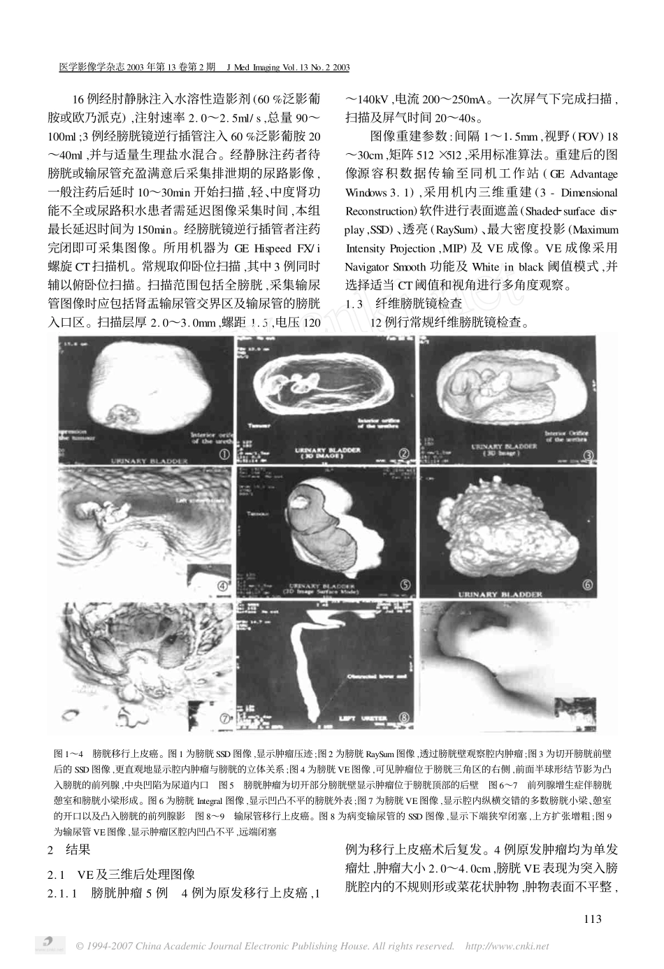
结果:膀胱肿瘤5例,前列腺增生8例(其中1例合并前列腺癌),输尿管狭窄4例,神经原性膀胱合并输尿管扩张1例。
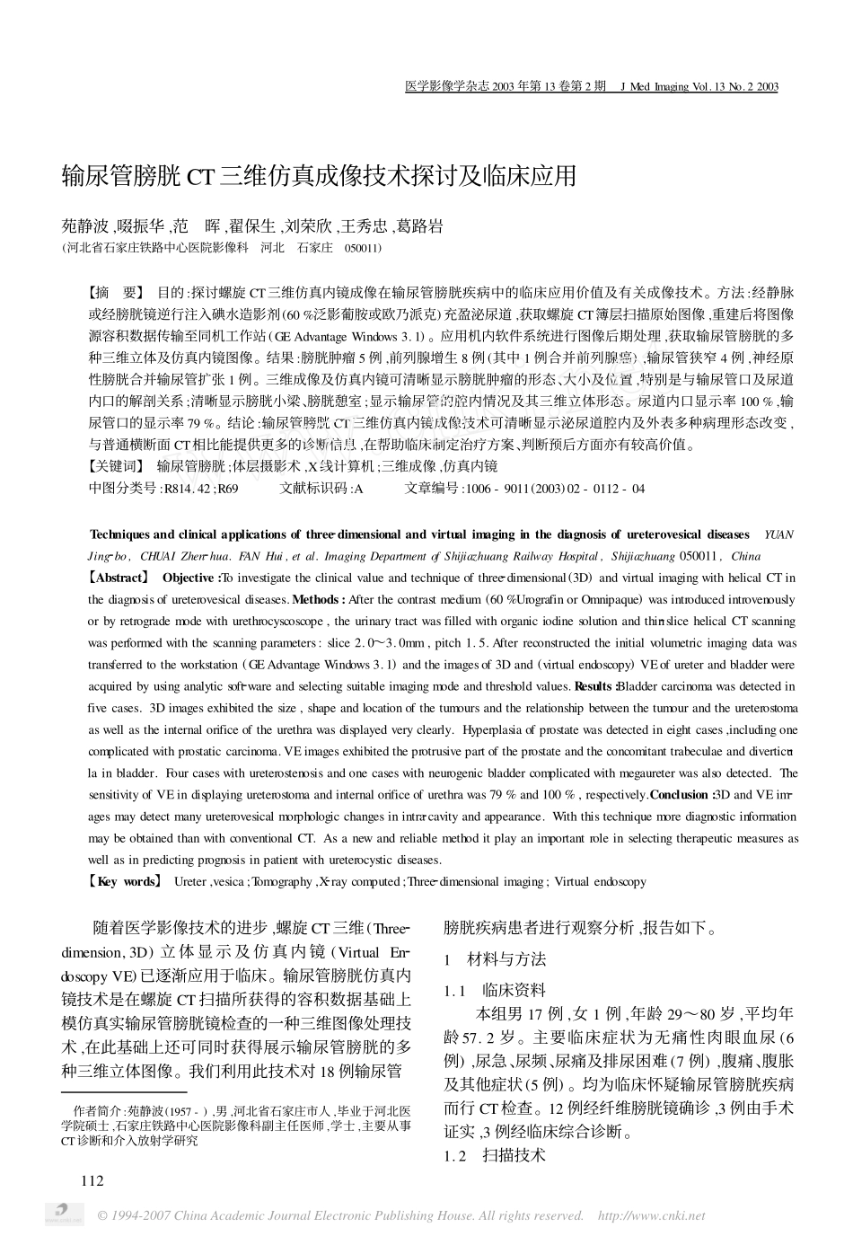
三维成像及仿真内镜可清晰显示膀胱肿瘤的形态、大小及位置,特别是与输尿管口及尿道内口的解剖关系;清晰显示膀胱小梁、膀胱憩室;显示输尿管的腔内情况及其三维立体形态。尿道内口显示率100%,输尿管口的显示率79%。结论:输尿管膀胱CT三维仿真内镜成像技术可清晰显示泌尿道腔内及外表多种病理形态改变,与普通横断面CT相比能提供更多的诊断信息,在帮助临床制定治疗方案、判断预后方面亦有较高价值。【关键词】输尿管膀胱;体层摄影术,X线计算机;三维成像,仿真内镜中图分类号:R814.42;R69文献标识码:A文章编号:1006-9011(2003)02-0112-04Techniquesandclinicalapplicationsofthree2dimensionalandvirtualimaginginthediagnosisofureterovesicaldiseasesYUANJing2bo,CHUAIZhen2hua.FANHui,etal.ImagingDepartmentofShijiazhuangRailwayHospital,Shijiazhuang050011,China【Abstract】Objective:Toinvestigatetheclinicalvalueandtechniqueofthree2dimensional(3D)andvirtualimagingwithhelicalCTinthediagnosisofureterovesicaldiseases.Methods:Afterthecontrastmedium(60%UrografinorOmnipaque)wasintroducedintrovenouslyorbyretrogrademodewithurethrocyscoscope,theurinarytractwasfilledwithorganiciodinesolutionandthin2slicehelicalCTscanningwasperformedwiththescanningparameters:slice2.0~3.0mm,pitch1.5.Afterreconstructedtheinitialvolumetricimagingdatawastransferredtotheworkstation(GEAdvantageWindows3.1)andtheimagesof3Dand(virtualendoscopy)VEofureterandbladderwereacquiredbyusinganalyticsoft2wareandselectingsuitableimagingmodeandthresholdvalues.Results:Bladdercarcinomawasdetectedinfivecases.3Dimagesexhibitedthesize,shapeandlocationofthetumoursandtherelationshipbetweenthetumourandtheureterostomaaswellastheinternalorificeoftheurethrawasdisplayedveryclearly.Hyperplasiaofprostatewasdetectedineightcases,includingonecomplicatedwithprostaticcarcinoma.VEimagesexhibitedtheprotrusivepartoftheprostateandtheconcomitanttrabeculaeanddiverticu2lainbladder.Fourcaseswithureterostenosisandonecaseswithneurogenicbladdercomplicatedwithmegaureterwasalsodetected.ThesensitivityofVEindisplayingureterostomaandinternalorificeofurethrawas79%and100%,respectively.Conclusion:3DandVEim2agesmaydetectmanyureterovesicalmorphologicchangesinintra2cavityandappearance.WiththistechniquemorediagnosticinformationmaybeobtainedthanwithconventionalCT.Asanewandreliablemethoditplayanimportantroleinselectingtherapeuticmeasuresaswellasinpredictingprognosisinpatientwithureterocysticdiseases.【Keywords】Ureter,vesica;Tomography,X2raycomputed;Three2dimensionalimaging;Virtualendoscopy随着医学影像技术的进步,螺旋CT三维(Three2dimension,3D)立体显示及仿真内镜(VirtualEn2doscopyVE)已逐渐应用于临床。输尿管膀胱仿真内镜技术是在螺旋CT扫描所获得的容积数据基础上模仿真实输尿管膀胱镜检查的一种三维图像处理技术,在此基础上还可同时获得展示输尿管膀胱的多种三维立体图像。我们利用此技术对18例输尿管作者简介:苑静波(1957-),男,河北省石家庄市人,毕业于河北医学院硕士,石家庄铁路中心医院影像科副主任...

1、当您付费下载文档后,您只拥有了使用权限,并不意味着购买了版权,文档只能用于自身使用,不得用于其他商业用途(如 [转卖]进行直接盈利或[编辑后售卖]进行间接盈利)。
2、本站所有内容均由合作方或网友上传,本站不对文档的完整性、权威性及其观点立场正确性做任何保证或承诺!文档内容仅供研究参考,付费前请自行鉴别。
3、如文档内容存在违规,或者侵犯商业秘密、侵犯著作权等,请点击“违规举报”。

碎片内容

输尿管膀胱CT三维仿真成像技术探讨及临床应用

确认删除?
VIP
微信客服
  • 扫码咨询
会员Q群
  • 会员专属群点击这里加入QQ群
客服邮箱
回到顶部