中国组织工程研究与临床康复第14卷第52期2010–12–24出版JournalofClinicalRehabilitativeTissueEngineeringResearchDecember24,2010Vol.14No.52P.O.Box1200,Shenyang110004cn.zglckf.com97641DepartmentofOtolaryngologyHeadandNeckSurgery,2DepartmentofComputedTomography,GeneralHospitalofChinesePeople'sArmedPoliceForces,Beijing100039,ChinaWangXiao-lu,☆Doctor,Attendingphysician,DepartmentofOtolaryngologyHeadandNeckSurgery,GeneralHospitalofChinesePeople'sArmedPoliceForces,Beijing100039,Chinalwh_ln@163.comCorrespondenceto:ShanXi-zheng,Master,Professor,DepartmentofOtolaryngologyHeadandNeckSurgery,GeneralHospitalofChinesePeople'sArmedPoliceForces,Beijing100039,ChinaSupportedby:agrantfromGeneralHospitalofChinesePeople'sArmedPoliceForces,No.
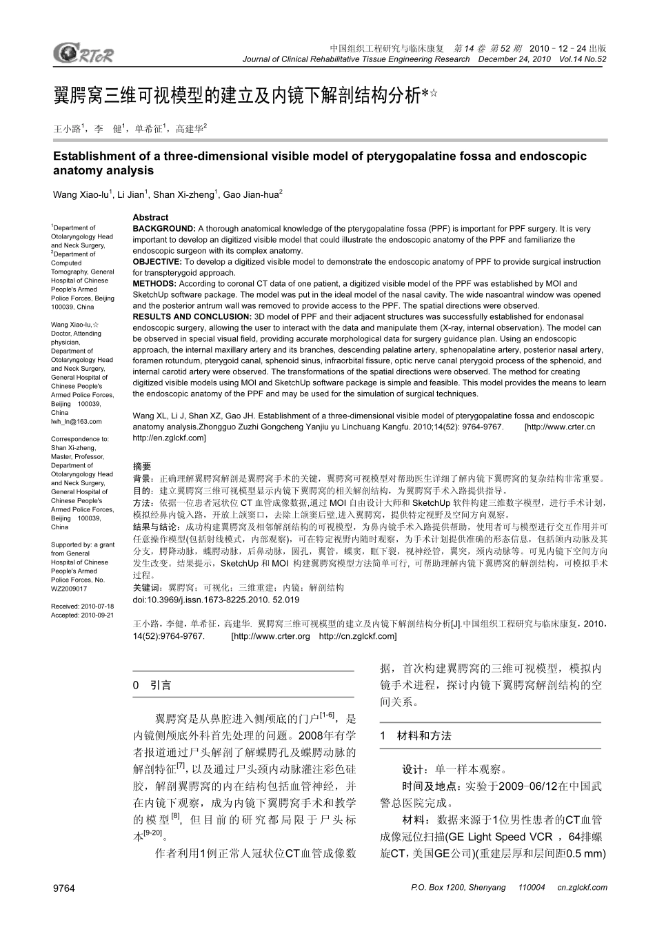
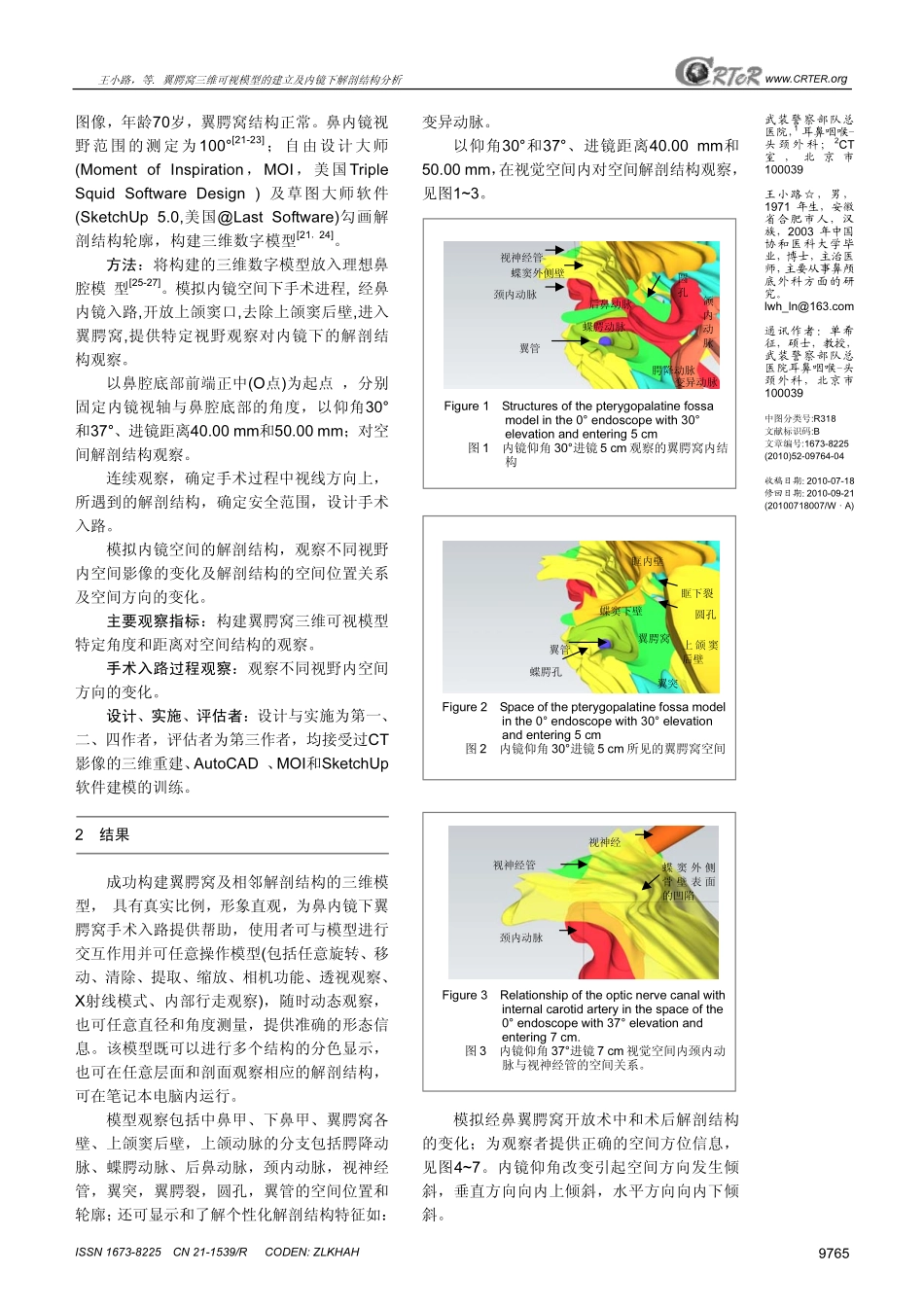
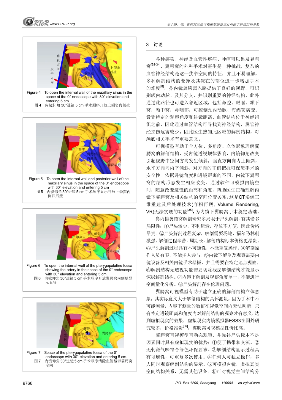
WZ2009017Received:2010-07-18Accepted:2010-09-21翼腭窝三维可视模型的建立及内镜下解剖结构分析*☆王小路1,李健1,单希征1,高建华2Establishmentofathree-dimensionalvisiblemodelofpterygopalatinefossaandendoscopicanatomyanalysisWangXiao-lu1,LiJian1,ShanXi-zheng1,GaoJian-hua2AbstractBACKGROUND:Athoroughanatomicalknowledgeofthepterygopalatinefossa(PPF)isimportantforPPFsurgery.ItisveryimportanttodevelopandigitizedvisiblemodelthatcouldillustratetheendoscopicanatomyofthePPFandfamiliarizetheendoscopicsurgeonwithitscomplexanatomy.OBJECTIVE:TodevelopadigitizedvisiblemodeltodemonstratetheendoscopicanatomyofPPFtoprovidesurgicalinstructionfortranspterygoidapproach.METHODS:AccordingtocoronalCTdataofonepatient,adigitizedvisiblemodelofthePPFwasestablishedbyMOIandSketchUpsoftwarepackage.Themodelwasputintheidealmodelofthenasalcavity.ThewidenasoantralwindowwasopenedandtheposteriorantrumwallwasremovedtoprovideaccesstothePPF.Thespatialdirectionswereobserved.RESULTSANDCONCLUSION:3DmodelofPPFandtheiradjacentstructureswassuccessfullyestablishedforendonasalendoscopicsurgery,allowingtheusertointeractwiththedataandmanipulatethem(X-ray,internalobservation).Themodelcanbeobservedinspecialvisualfield,providingaccuratemorphologicaldataforsurgeryguidanceplan.Usinganendoscopicapproach,theinternalmaxillaryarteryanditsbranches,descendingpalatineartery,sphenopalatineartery,posteriornasalartery,foramenrotundum,pterygoidcanal,sphenoidsinus,infraorbitalfissure,opticnervecanalpterygoidprocessofthesphenoid,andinternalcarotidarterywereobserved.Thetransformationsofthespatialdirectionswereobserved.ThemethodforcreatingdigitizedvisiblemodelsusingMOIandSketchUpsoftwarepackageissimpleandfeasible.ThismodelprovidesthemeanstolearntheendoscopicanatomyofthePPFandmaybeusedforthesimulationofsurgicaltechniques.WangXL,LiJ,ShanXZ,GaoJH.Establishmentofathree-dimensionalvisiblemodelofpterygopalatinefossaandendoscopicanatomyanalysis.ZhongguoZuzhiGongchengYanjiuyuLinchuangKangfu.2010;14(52):9764-9767.[http://www.crter.cnhttp://en.zglckf.com]摘要背景:正确理解翼腭窝解剖是翼腭窝手术的关键,翼腭窝可视模型对帮助医生详细了解内镜下翼腭窝的复杂结构非常重要。目的:建立翼腭窝三维可视模型显示内镜下翼腭窝的相关解剖结构,为翼腭窝手术入路提供指导。方法:依据一位患者冠状位CT血管成像数据,通过MOI自由设计大师和SketchUp软件构建三维数字模型,进行手术计划,模拟经鼻内镜入路,开放上颌窦口,去除上颌窦后壁,进入翼腭窝,提供特定视野及空间方向观察。结果与结论:成功构建翼腭窝及相邻解剖结构的可视模型,为鼻内镜手术入路提供帮助,使用者可与模型进行交互作用并可任意操作模型(包括射线模式,内部观察),可在特定视野内随时观察,为手术计划提供准确的形态信息,包...