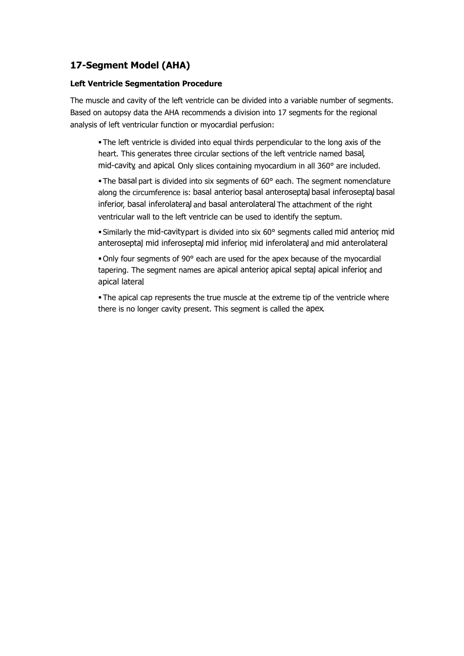
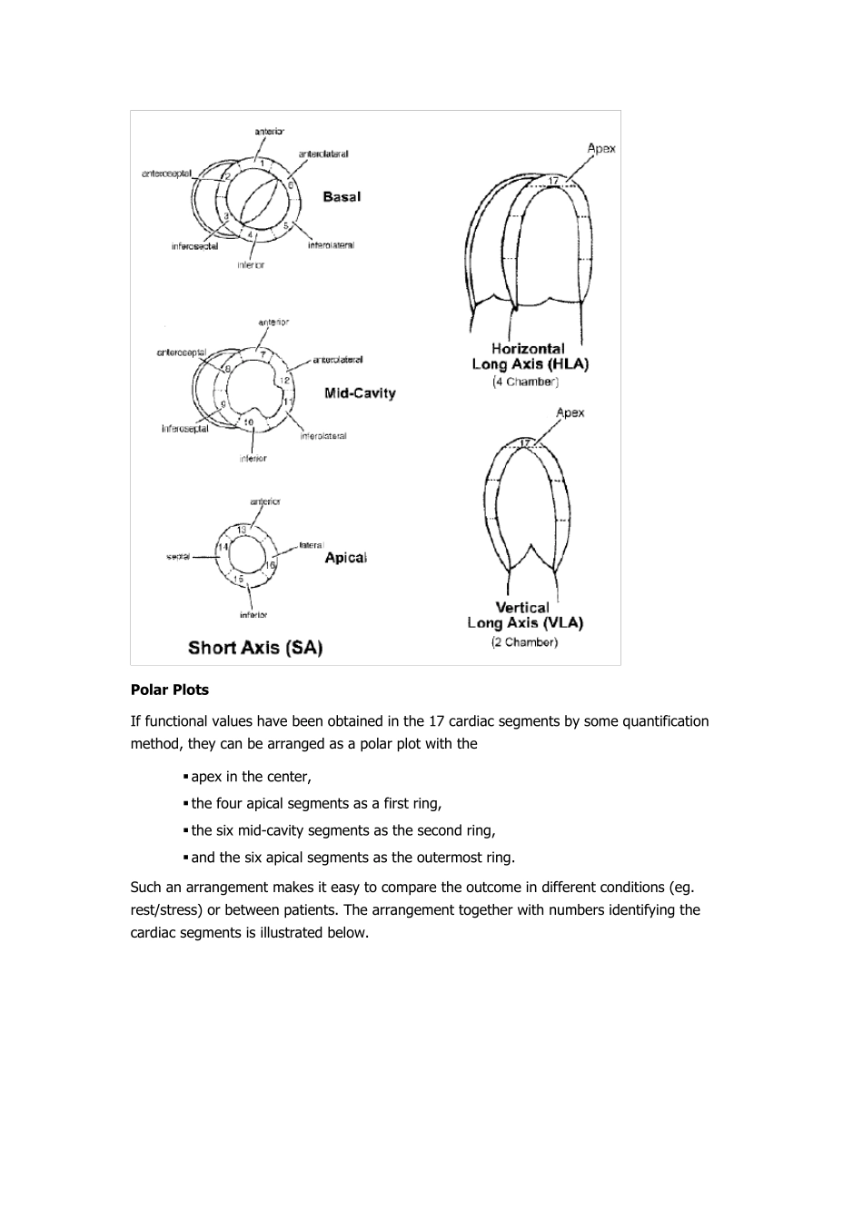
17-Segment Model (AHA) Left Ventricle Segmentation Procedu re The muscle and cavity of the left ventricle can be divided into a variable number of segments. Based on autopsy data the AHA recommends a division into 17 segments for the regional analysis of left ventricular function or myocardial perfusion: ▪ The left ventricle is divided into equal thirds perpendicular to the long axis of the heart. This generates three circular sections of the left ventricle named basal, mid-cavity, and apical. Only slices containing myocardium in all 360° are included. 
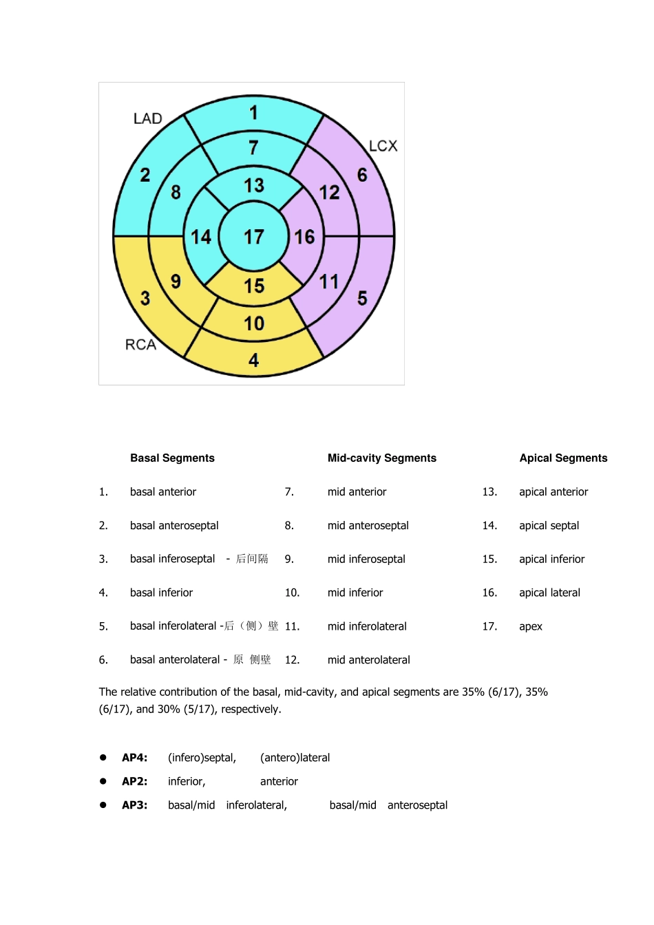
▪ The basal part is divided into six segments of 60° each. The segment nomenclature along the circumference is: basal anterior, basal anteroseptal, basal inferoseptal, basal inferior, basal inferolateral, and basal anterolateral. The attachment of the right ventricular wall to the left ventricle can be used to identify the septum. ▪ Similarly the mid-cavity part is divided into six 60° segments called mid anterior, mid anteroseptal, mid inferoseptal, mid inferior, mid inferolateral, and mid anterolateral. ▪ Only four segments of 90° each are used for the apex because of the myocardial tapering. The segment names are apical anterior, apical septal, apical inferior, and apical lateral. ▪ The apical cap represents the true muscle at the extreme tip of the ventricle where there is no longer cavity present. This segment is called the apex. Polar Plots If functional values have been obtained in the 17 cardiac segments by some quantification method, they can be arranged as a polar plot with the ▪ apex in the center, ▪ the four apical segments as a first ring, ▪ the six mid-cavity segments as the second ring, ▪ and the six apical segments...