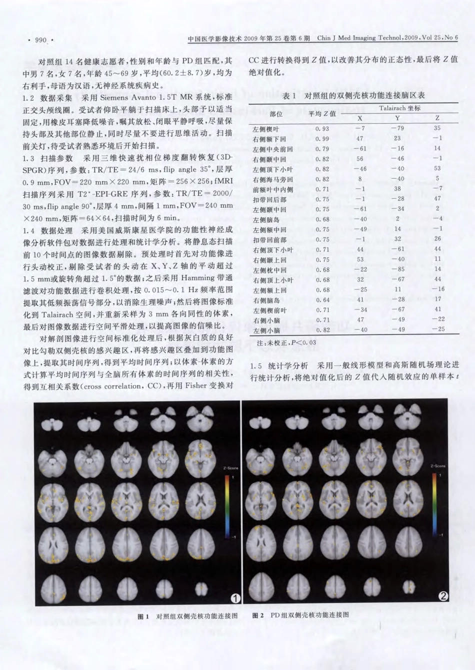
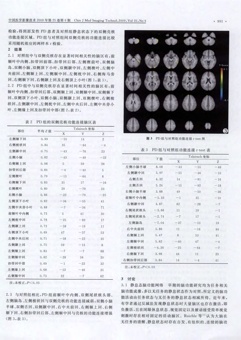
中国医学影像技术2009年第25卷第6期ChinJMedImagingTechnol,2009,Vol25,No6·989·◆:◆中枢神经影像学FunctionalMRIinvestigationofbrainfunctionalconnectivityinresting—stateofParkinson’SdiseasepatientsCHENJun,LIUBo,LIUXian,CHENZhi—guang,LINing—na,LUOXiao—dong(1.DepartmentofRadiology,2.DepartmentofNeurology,GuangdongProvincialTraditionalChineseMedicalHospital,Guangzhou510120,China)~Abstract]ObjectiveToinvestigatethecharacteristicsofbrainfunctionalconnectivityinresting—stateofParkinson’Sdis—ease(PD)patientswithfunctionalMRI(fMRI).MethodsFourteenPDpatients(PDgroup)and14matchedcontrolsun—derwentexaminationinresting—statewithfMRI.Putamensofbilateralweretheregionsofinterest,andthedifferenceofPDpatientsbetweencontrolswasanalyzedwithtwo—samplettest.ResultsComparedwithcontrols,functionalconnectivityinresting—stateofPDpatientswasabnormal,includingdecreasedfunctionalconnectivityandincreasedfunctionalconnectivity.Thebrainareaofdecreasedfunctionalconnectivityincludedmedialprefrontalcortex,bilateralcaudatehead,leftinsula,leftfusiformgyrus.Thebrainareaofincreasedfunctionalconnectivityincludedbilateralcerebellartonsil,bilaterallingualgyrus,bilateralmiddletemporalgyrus,rightprecentralgyrus,rightsuperiorfrontalgyrus,rightinferiorfrontalgyrus,rightein—gulategyrusandleftmiddlefrontalgyrus.ConclusionThefunctionalconnectivityofPDpatientsisabnormalinrestingState.[Keywords]Parkinsondisease;Resting-state;Functionalmagneticresonanceimaging功能磁共振成像评价帕金森患者静息状态下脑功能连接陈俊,刘波H,刘岘,陈志光,李宁娜,雒晓东(1.广东省中医院影像科;2.神经内科,广东广州510120)[摘要]目的采用功能磁共振成像(fMRI)评价帕金森病(PD)患者静息状态下脑功能连接的特点。方法对14例符合PD诊断标准的患者及14名健康者行静息态脑fMRI,选双侧壳核为感兴趣区,PD患者与正常对照组间脑功能连接的差异用随机效应两样本t检验。结果与正常对照组相比,PD患者在静息状态下前额叶中内侧、双侧尾状核头部、左侧脑岛、左侧梭状回等脑区功能连接减低;双侧小脑半球、双侧舌回、双侧颞中回、右中央前回、右侧额上回、右侧额下回、右侧扣带回后部、左侧额中回等脑区功能连接增强。结论PD患者在静息状态下存在异常的脑功能连接网络。[关键词]帕金森病;静息状态;功能磁共振成像[中图分类号]R742.5;R445.2[文献标识码]A[文章编号]1003—3289(2009)06—0989—04帕金森病(Parkinson’Sdisease,PD)是人类最常见的神经退行性疾病之一。静息态脑功能磁共振成像(functionalmagneticresonanceimaging,fMRI)是近年发展的一种新的脑功能成像方法,已经应用于阿尔茨海默病、幻听等疾病的早期研究。本研究对早中期PD患者的静息态脑fMRI进行[基金项目]广东省科技计划项目(2008B080703041)。[作者简介]陈俊(1981一),男,广东五华人,本科,医师。研究方向:磁共振脑功能成像。E—mail:junesums@163.corn[通讯作者]刘波.广州中医药大学附属第二医院,510I20。Email:Doctorlb2006@yahoo.corn.cn[收稿日期]20080818[修回日期]20090224分析。1资料与方法1.1研究对象参照《2006年中华医学会神经病学分会运动障碍及帕金森病学组帕金森病诊断指南》,选取2007年1—12月在我院帕金森专科门诊就诊的14例原发性PD患者(PD组),排除各种原因引起的继发性PD和帕金森叠加综合征。所有患者均为右利手,母语为汉语,临床症状均为右侧起病,其中男7例,女7例,年龄45~70岁,平均(60.1_4-8.9)岁,Hoehn-Yahr分级Ⅱ级6例,Ⅲ级8例,PD综合评分分数为(29.2±5.2)分。14例PD患者中有6例正在接受药物治疗,余8例均未接受药物治疗;所有正在接受治疗者检查前抗PD药物至少停用12h。990中国医学影像技术2009年第25卷第6期ChinJMedImagingTeehnol,2009,Vol25...