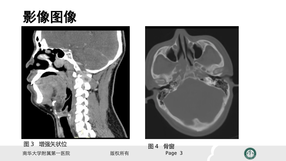
电脑桌面
添加小米粒文库到电脑桌面
安装后可以在桌面快捷访问

鼻咽纤维血管瘤的影像表现及临床VIP免费

鼻咽纤维血管瘤的影像表现及临床_第1页
1/18
鼻咽纤维血管瘤的影像表现及临床_第2页
2/18
鼻咽纤维血管瘤的影像表现及临床_第3页
3/18
读书报告会鼻咽纤维血管瘤的影像表现及临床南华大学附属第一医院版权所有Page2患者:男,26岁主诉:右鼻出血2天图1CT平扫图2CT增强南华大学附属第一医院版权所有影像图像Page3图3增强矢状位图4骨窗南华大学附属第一医院版权所有影像图像Page4图5MRIT1WI图6MRIT2WI南华大学附属第一医院版权所有影像图像Page5图7MRIT1WI增强图8MRIT1WI增强图9MRIT1WI增强南华大学附属第一医院版权所有影像图像Page6图10DSA冠状位图11DSA矢状位南华大学附属第一医院版权所有Page7患者:男,26岁主诉:右鼻出血2天现病史:患者输2天前无明显诱因出现右鼻出血,为鲜血,呈滴状,先从左前鼻孔出,后亦从口中、右鼻流出,数分钟后停止,反复出现多次,总量约为100ml,无鼻塞,流涕,嗅觉正常。无头痛、发热、咳嗽、打鼾,无耳鸣、而鼻塞感,无听力下降。于当地医院治疗,予以鼻腔填塞,症状好转。在中山陈星海医院,予以电子喉镜检查“右鼻腔肿物,性质待查”。既往史:否认肝炎、结核、疟疾病史,否认高血压、心脏病史,否认糖尿病、脑血管疾病史,否认手术、外伤、输血史,否认食物、药物等过敏史,否认吸烟、饮酒史,否认毒物接触史。南华大学附属第一医院版权所有AbstractNasopharyngealangiofibroma(NA)isarare,vasculartumoraffectingdolescentmales.Duetoaggressivelocalgrowth,skullbaselocationandriskofprofoundhemorrhage,NAisachallengeforsurgeons.AngiofibromastumorshowedintensivecontrastenhancementonCTandmagneticresonanceimaging(MRI)scans,andabundantvascularityonangiography.Page8南华大学附属第一医院版权所有Background(NA)isararevasculartumor,whichrepresents0.05%ofallheadandnecktumors.Atthesametime,itisthemostcommonbenignneoplasmofthenasopharynx.NAoccurspredominantlyinadolescentmales.Althoughhistologicallybenignitshowslocallyaggressivegrowthwithbonedestructionandspreadthroughnaturalforaminaandfissures.Page9南华大学附属第一医院版权所有Itoriginatesfromtheposterolateralwallofthenasopharynxandfromthissiteusuallyextendstothenasopharynx,nasalcavity,paranasalsinuses,sphenoid-palatineforamenandinfratemporalfossa.In10–20%ofthecasestumorinvadesthecranialcavity。Page10南华大学附属第一医院版权所有NasaltumorunderwentCT,whichdemonstratedhomogenousmass,withcontrastenhancementrangingfromstrongtointermediate(Fig.1).Inonecase,signsofbonydestructionwithtumorinvasiontotheethmoidsinuswerevisible.ThepatientwiththetumoroftheinfratemporalfossaunderwentCT,(MRI)andcarotidarteriographywithpreoperativeembolization.Thelesionshowedintensivecontrast。Page11南华大学附属第一医院版权所有Page12Fig.1Computedtomography,coronalplane,showshomogenoustumormassintherightnasalcavityFig.2Magneticresonance,saggitalT1-weightedimageaftercontrastadministration.南华大学附属第一医院版权所有Page13Histologicsectionofthetumor(H&Estain)showsfibrousstromawithectatic,thin-walledvascularchannelsEnhancementonCTandMRIaswellassignal-voidareasonMRimages,typicalforhighflowvessels(Fig.2).Arteriographyrevealedabundantvascularitywithmainbloodsupplyfromtheinternalmaxillaryartery.南华大学附属第一医院版权所有EnhancementonCTandMRIaswellassignal-voidareasonMRimages,typicalforhighflowvessels(Fig.2).Arteriographyrevealedabundantvascularitywithmainbloodsupplyfromtheinternalmaxillaryartery.Page14南华大学附属第一医院版权所有HistopathologicalappearancetypicalforNAconsistsofnumerouswide,irregularvesselswithasinglelayerofendothelialcells,embeddedinfibrousstroma.Theabundantvascularcomponentisresponsibleforexcessivebleedingduringsurgeryorfollowingbiopsies.ItalsocontributestocertaincharacteristicradiologicalfeaturesofNAs,includingstrongcont...

1、当您付费下载文档后,您只拥有了使用权限,并不意味着购买了版权,文档只能用于自身使用,不得用于其他商业用途(如 [转卖]进行直接盈利或[编辑后售卖]进行间接盈利)。
2、本站所有内容均由合作方或网友上传,本站不对文档的完整性、权威性及其观点立场正确性做任何保证或承诺!文档内容仅供研究参考,付费前请自行鉴别。
3、如文档内容存在违规,或者侵犯商业秘密、侵犯著作权等,请点击“违规举报”。

碎片内容

鼻咽纤维血管瘤的影像表现及临床

您可能关注的文档

确认删除?
VIP
微信客服
  • 扫码咨询
会员Q群
  • 会员专属群点击这里加入QQ群
客服邮箱
回到顶部