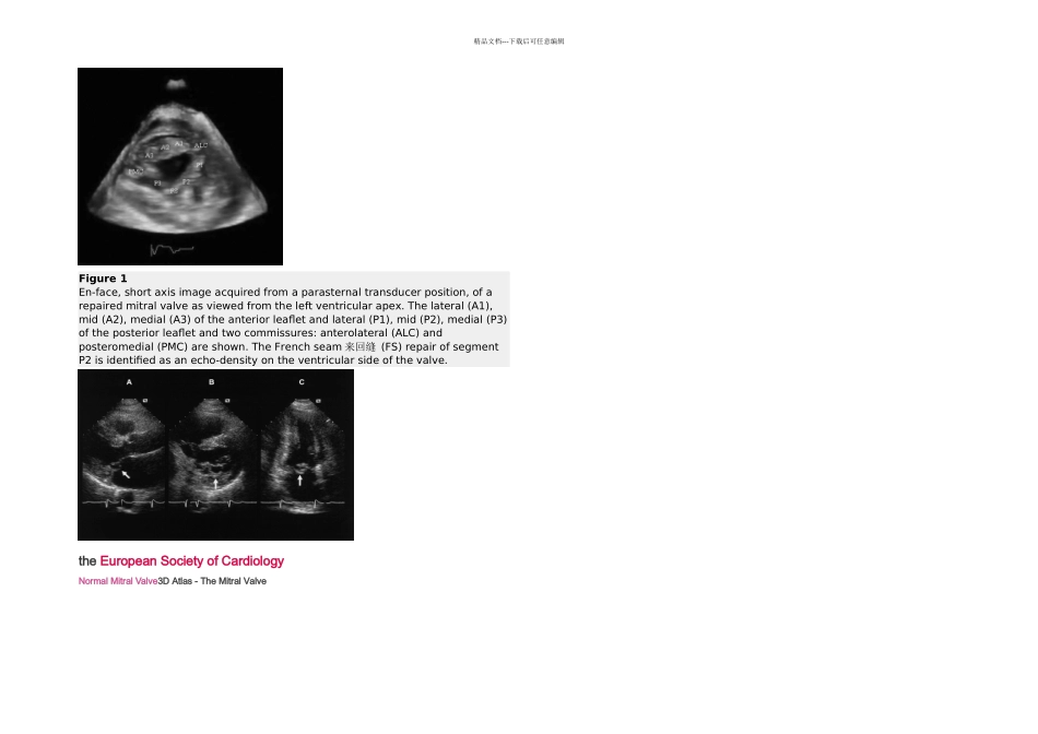
精品文档---下载后可任意编辑Figure 1En-face, short axis image acquired from a parasternal transducer position, of a repaired mitral valve as viewed from the left ventricular apex
The lateral (A1), mid (A2), medial (A3) of the anterior leaflet and lateral (P1), mid (P2), medial (P3) of the posterior leaflet and two commissures: anterolateral (ALC) and posteromedial (PMC) are shown
The French seam 来回缝 (FS) repair of segment P2 is identified as an echo-density on the ventricular side of the valve
the European Society of CardiologyNormal Mitral Valve3D Atlas - The Mitral Valve