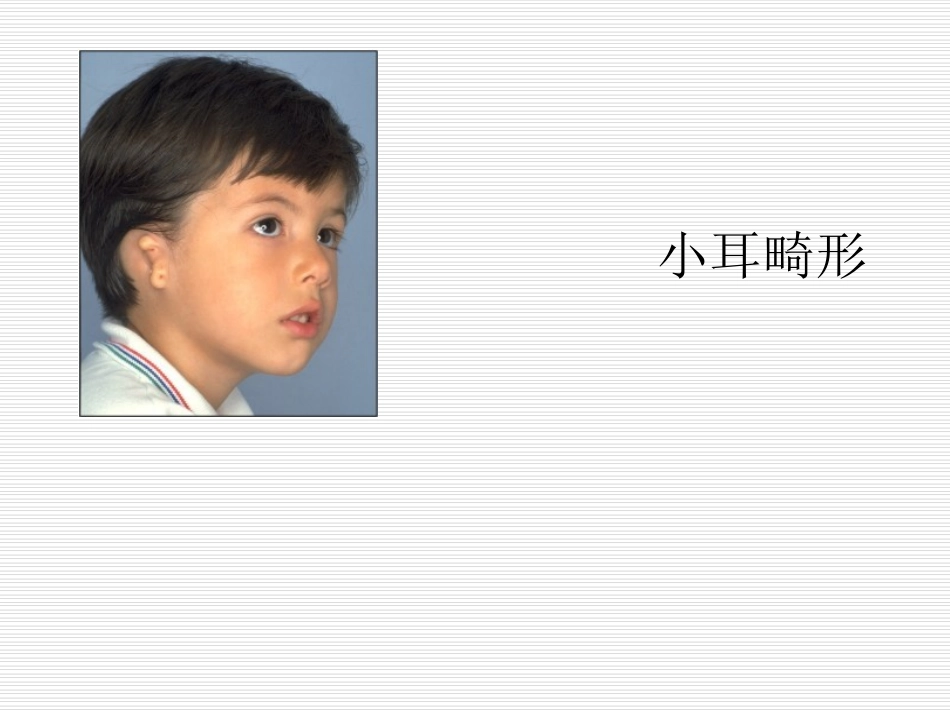
小耳畸形TheNagataTechnique•BackgroundFirstintroducedin1993,theNagatatechniquehasenjoyedwidesuccessasanalternativetotheBrenttechnique.Itsmajoradvantageliesinitstwo-stagedapproach•ThefirststageoftheNagatatechniqueinvolves:1.Fabricationandinsertionofacartilageframework2.TranspositionofthelobuleThisroughlycorrespondstothefirstthreestagesoftheBrenttechniqueFirstStageUsetheipsilateral6th–9thcostalcartilagesinfabricatingtheframeworkHarvestingofthecostalcartilages•Theframeworkisconstructedinthreedistinctlevelsor“floors”1.Firstfloor:thecrushelicis、fossatriangularis2.Secondfloor:thescapha3.Thirdfloor:thehelix、antihelix、tragus,antitragusFabrication–The6thand7thisbaseframe–The8thisthehelixandcrushelicis–The9thisthesuperiorcrus,inferiorcrus、andantihelixInsertthecartilageframework1.A“W”incisiononlobuleremnant2.TheskinflapiselevatedtoreceivetheframeworkBolstersofdentalcottonarethenusedtosecuretheskinflapstothecartilageframework.Theseareaffixedwith4-0monofilamentmattresssutures.Thebolstersarekeptinplacefor2weeks3.InsertthecartilageframeworkSecondStageElevatetheconstructionSecondStageThetemporoparietalfascialflapElevatetheconstructionRaisingtherotationflapforcoveringtheadditionalcartilagegraftforprotrusionoftheauricleFlaptranspositionforcoverageofthecochalwallcartilagegraftRotationflapcoverscochalwallcartilagegraft.Splitthicknessscapleskingraftcoverstheposterioraspectoftheelevatedauriclebeyondthecochalwallgraft.Donorsiteoftheskingraftcoveredwithvaselinegauze.Posteriorview.Earlypost-operativeresult(10days).Lateralview(10days)6monthspost-op扩张器植入及注液扩张于耳后发际内1cm处设计平行于发际切口,长约3~5cm,在颞肌浅筋膜上、胸锁乳突肌腱膜上和残耳软骨与软骨膜问进行潜行分离囊袋.植入扩张器,注射壶植入颈部皮下。扩张器法耳再造第一期术后第8天开始注水.每次注射生理盐水3~8ml,每周注水3次.50ml扩张器可注水55~65ml。注水完毕1-2个月后行耳再造术。2monthsafterthefirstoperation软骨支架制备:切取第6-8肋软骨,根据健耳胶片模型、实际尺寸的患耳片(健耳镜面像),以及健耳外耳轮到颅侧皮肤的垂直距离尽量整体雕刻耳支架。软骨的拼接用记忆合金丝或细丝线。第二期软骨支架植入取出扩张器aftertheexpanderwasemoved,ananteriorlybasedexpandedskinflapwasshapedandanipsilateraltemporoparietalfasciaflapmeasuring10X10cmwasharvested制作蒂在前面的扩张皮瓣及耳后筋膜瓣祛除扩张皮瓣浅层纤维包膜.使皮肤变薄,将软骨支架置入皮瓣和筋膜瓣之间固定残耳向后下转位形成耳垂.支架的前面覆盖扩张皮瓣。耳后创面植皮,打包包扎,负压引流管5d拔除,10d拆线。耳甲腔成形及部分残畸软骨切除耳再造4个月后,于耳甲腔处设计一“C”型皮瓣向前推进折叠形成耳屏,切除残畸软骨及多余的软组织.修整高起的残耳皮肤,尽量带蒂转移覆盖耳甲腔.去除耳甲腔内多余的软组织,直达乳突表面。创面植皮,打包包扎,10d拆线。第三期聚乙烯塑胶Medpor其优点除了可以避免切取肋软骨造成的创伤外,还可以规避术者雕刻技术欠佳的弱点,但高外露率是制约Medpor应用的瓶颈。早期的外露主要与颞浅筋膜的血供有关,远期外露主要与患者自身的保护、护理以及材料本身的因素有关。组织工程