��项目来源:国家重点基础研究发展计划资助(2010CB530507)、国家自然科学基金(30672593)、北京市自然科学基金(7062049)通讯作者:景向红,博士,研究员,主要从事针灸作用机制研究。Tel:(010)64014411-2750�实验研究�肥大细胞和P物质参与急性胃黏膜损伤大鼠体表穴位的敏化过程石�宏�程�斌1�李江慧�陈淑莉�谭奇纹1�晋志高�景向红(中国中医科学院针灸研究所经络研究中心,北京100700;1山东中医药大学,济南250355)摘�要!
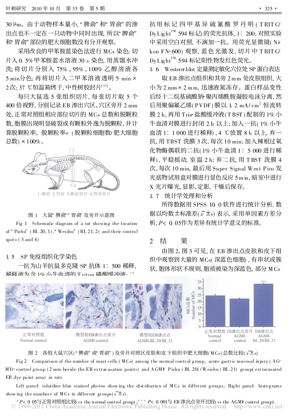
�目的:观察急性胃黏膜损伤后伊文思蓝(EB)渗出穴区肥大细胞聚集数量和脱颗粒变化及其局部P物质(SP)的表达,以揭示病理状态下穴位的组织细胞特征,确定肥大细胞和SP参与穴位敏化的过程。方法:大鼠随机分为模型组和正常对照组,每组15只。采用空腹稀盐酸灌胃造成急性胃黏膜损伤模型。5h后尾静脉注射EB,模型组取EB渗出点∀脾俞#∀胃俞#和旁开2mm处的皮肤和皮下组织,正常对照组取对应点皮肤和皮下组织。采用甲苯胺蓝染色法标记这些部位肥大细胞的聚集分布数量及脱颗粒特征。采用免疫组织化学方法显示局部SP的分布并进行Westernblot相对定量测定。结果:急性胃黏膜损伤后EB体表穴区渗出点的皮肤和皮下组织中肥大细胞呈现聚集,其数量和脱颗粒数明显多于正常对照组和∀脾俞#∀胃俞#旁开对照组(P<0�05,P<0�001),脱颗粒率也显著高于旁开点和正常对照组(P<0�01)。在EB渗出点和旁开部位均可见SP蛋白的表达水平显著高于正常组(P<0�01),其阳性纤维多分布于皮下。结论:在急性胃黏膜损伤时,体表出现的敏化穴位中肥大细胞参与了与疾病相关的体表穴位敏化过程,且其局部的SP表达水平增高。关键词!�急性胃黏膜损伤�敏化穴位�神经源性炎性反应�肥大细胞�P物质中图分类号!R224�2�文献标志码!A�文章编号!1000�0607(2010)05�0323�07MastCellandSubstancePAreInvolvedintheProcessofAcupointSensitizationInducedbyA�cuteGastricMucosalInjurySHIHong,�CHENGBin1,�LIJiang�hui,�CHENShu�li,�TANQi�wen1,�JINZhi�gao,�JINGXiang�hong(InstituteofAcu�moxibustion,ChinaAcademyofChineseMedicalSciences,Beijing100700,China;1ShandongUniversityofTraditio�nalChineseMedicine,Jinan250355)ABSTRACT!�Objective�Toobservechangesofmastcells(MCs)numberandmorphology,andsubstanceP(SP)ex�pressioninEvansblue(EB)extravasatedregionaroundacupoint∀Pishu#(BL20)and∀Weishu#(BL21)afteracutegastricmuco�salinjury(AGMI)soastoinvestigatethemechanismunderlyingvisceralproblems�inducedacupointactivation.Methods�Thirtya�dultWistarratswererandomlydividedintonormalcontrol(n=15)andAGMIgroups(n=15).AGMImodelwasduplicatedbyperfusingtheratswith0�5mol/LHCl(1mL/100g)afterfastingfor20h.FivehoursafterAGMI,theratsweretreatedbytail�in�travenousinjectionofEBdye(5mg/100g,50mg/mLinnormalsaline)forinducingdye�plasmaextravasationintheskinaroundBL20,BL21regions,etc.attheback.Theratsofthenormalcontrolgroupweretreatedwithtail�intravenousinjectionof0�9%NaCl.Theskinandsubcutaneoustissues(2mm∃2mm)ofextravasatedEBdyepoints(BL20orBL21region)andthose2mmlateraltotheextravasatedEBdyepointsinthemodelgroupandthecorrespondingpointsinthenormalcontrolgroupweresampled(followedbyfixingthemin4%paraformaldehyde),sectionedandstainedbytoluidineblue(forlabelingMCs).TheexpressionofSPintheextravasatedEBdyeskinandsubcutaneoustissueswasdetectedbyimmunohistochemistry(n=5)andwesternblot(n=5)respectively.ThenumberofMCsinthesesampleswascountedandthedegranulationrateofMCscalculated.Results�ThetotalnumberofMCsandthenumberofdegranulatedMCsweresignificantlymoreintheEBextravasationpoints(correspon�dingtoBL20/BL21area)ofAGMIgroupthanthoseinthecontrolspotsofAGMIgroupandthanthoseinthenormalcontrolgroup%323%针刺研究�2010年10月�第35卷�第5期(P<0�05,P<0�001).Thedegranulat...