ChinComputMedImag2006,12:92~95中国医学计算机成像杂志2006,12:92~95中国医学计算机成像杂志2006年第12卷第2期-92-心脏原发恶性肿瘤CT及MRI表现杨姗张志勇曾蒙苏王佩芬陈财忠沈继章陈刚目的:了解心脏原发恶性肿瘤的CT、MRI表现,旨在提高诊断及鉴别诊断。材料和方法:12例经病理证实心脏恶性肿瘤的CT及MRI资料进行回顾性分析。其中5例行胸部CT平扫加增强扫描。9例做心脏MR检查,其中7例行动态增强扫描。
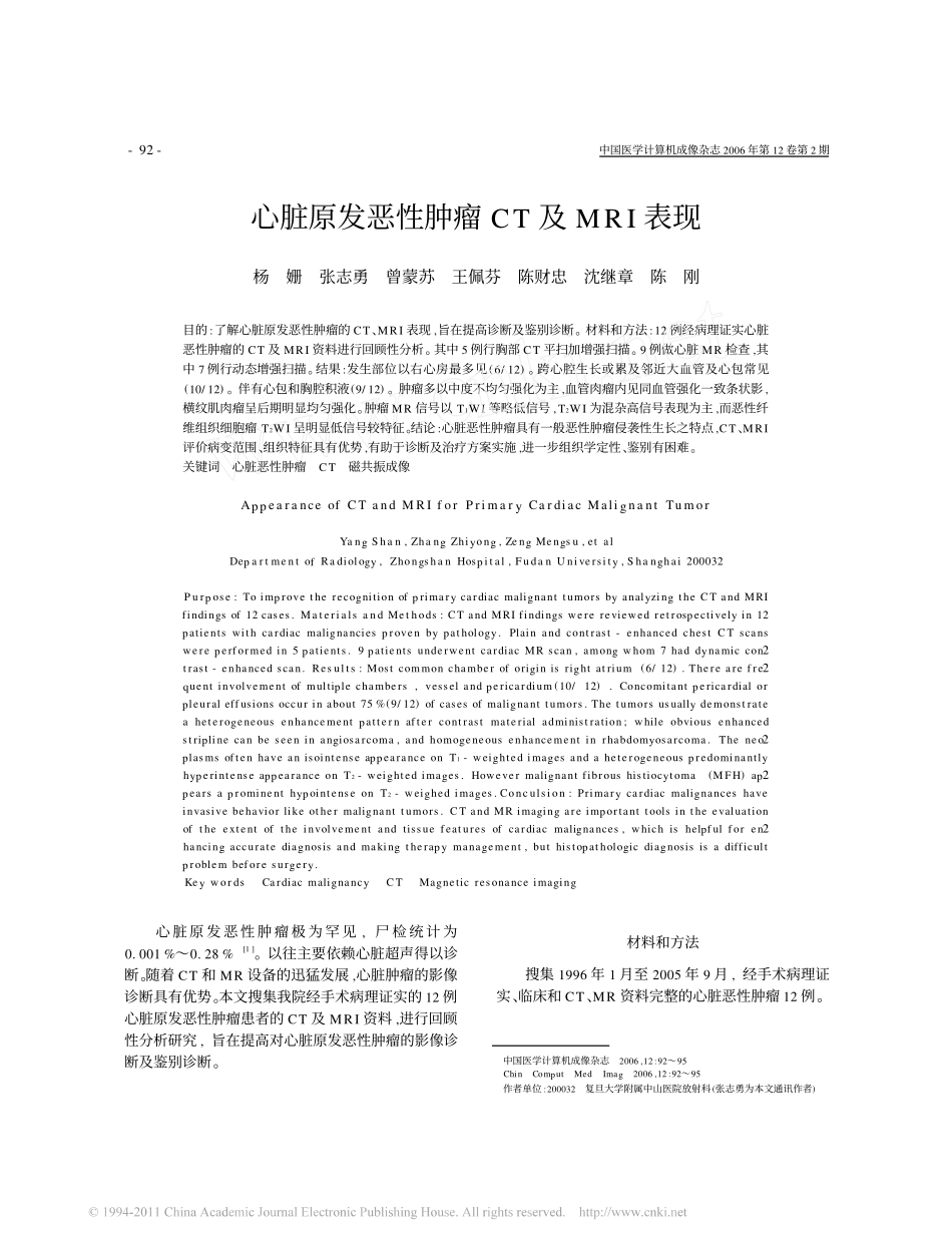
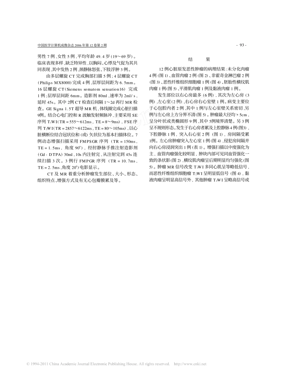
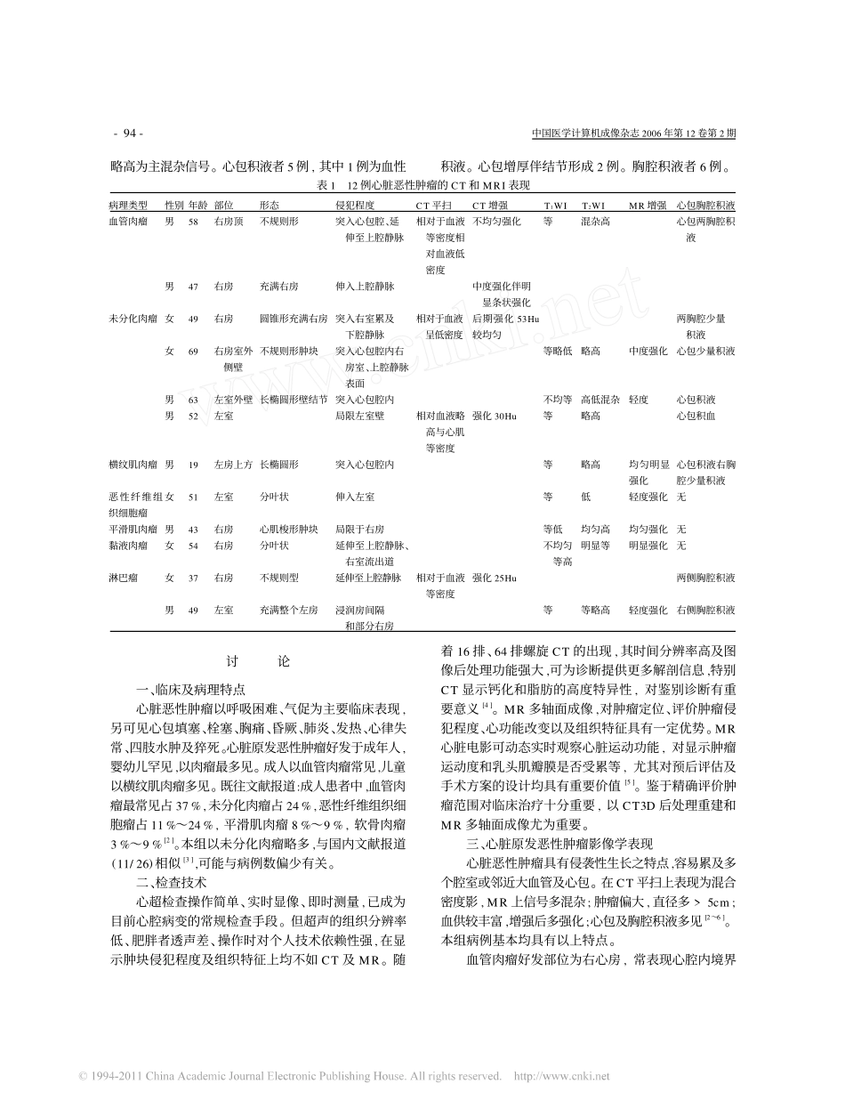
结果:发生部位以右心房最多见(6/12)。跨心腔生长或累及邻近大血管及心包常见(10/12)。伴有心包和胸腔积液(9/12)。肿瘤多以中度不均匀强化为主,血管肉瘤内见同血管强化一致条状影,横纹肌肉瘤呈后期明显均匀强化。肿瘤MR信号以T1WI等略低信号,T2WI为混杂高信号表现为主,而恶性纤维组织细胞瘤T2WI呈明显低信号较特征。结论:心脏恶性肿瘤具有一般恶性肿瘤侵袭性生长之特点,CT、MRI评价病变范围、组织特征具有优势,有助于诊断及治疗方案实施,进一步组织学定性、鉴别有困难。关键词心脏恶性肿瘤CT磁共振成像AppearanceofCTandMRIforPrimaryCardiacMalignantTumorYangShan,ZhangZhiyong,ZengMengsu,etalDepartmentofRadiology,ZhongshanHospital,FudanUniversity,Shanghai200032Purpose:ToimprovetherecognitionofprimarycardiacmalignanttumorsbyanalyzingtheCTandMRIfindingsof12cases.MaterialsandMethods:CTandMRIfindingswerereviewedretrospectivelyin12patientswithcardiacmalignanciesprovenbypathology.Plainandcontrast-enhancedchestCTscanswereperformedin5patients.9patientsunderwentcardiacMRscan,amongwhom7haddynamiccon2trast-enhancedscan.Results:Mostcommonchamberoforiginisrightatrium(6/12).Therearefre2quentinvolvementofmultiplechambers,vesselandpericardium(10/12).Concomitantpericardialorpleuraleffusionsoccurinabout75%(9/12)ofcasesofmalignanttumors.Thetumorsusuallydemonstrateaheterogeneousenhancementpatternaftercontrastmaterialadministration;whileobviousenhancedstriplinecanbeseeninangiosarcoma,andhomogeneousenhancementinrhabdomyosarcoma.Theneo2plasmsoftenhaveanisointenseappearanceonT1-weightedimagesandaheterogeneouspredominantlyhyperintenseappearanceonT2-weightedimages.Howevermalignantfibroushistiocytoma(MFH)ap2pearsaprominenthypointenseonT2-weighedimages.Conculsion:Primarycardiacmalignanceshaveinvasivebehaviorlikeothermalignanttumors.CTandMRimagingareimportanttoolsintheevaluationoftheextentoftheinvolvementandtissuefeaturesofcardiacmalignances,whichishelpfulforen2hancingaccuratediagnosisandmakingtherapymanagement,buthistopathologicdiagnosisisadifficultproblembeforesurgery.KeywordsCardiacmalignancyCTMagneticresonanceimaging心脏原发恶性肿瘤极为罕见,尸检统计为0.001%~0.28%[1]。以往主要依赖心脏超声得以诊断。随着CT和MR设备的迅猛发展,心脏肿瘤的影像诊断具有优势。本文搜集我院经手术病理证实的12例心脏原发恶性肿瘤患者的CT及MRI资料,进行回顾性分析研究,旨在提高对心脏原发恶性肿瘤的影像诊断及鉴别诊断。材料和方法搜集1996年1月至2005年9月,经手术病理证实、临床和CT、MR资料完整的心脏恶性肿瘤12例。作者单位:200032复旦大学附属中山医院放射科(张志勇为本文通讯作者)中国医学计算机成像杂志2006年第12卷第2期-93-男性7例,女性5例,平均年龄49.4岁(19~69岁)。临床表现多样,缺乏特异性,以胸闷、心悸及气促为其共同表现,其中发热2例,颈静脉怒张、下肢浮肿3例。由多层螺旋CT完成胸部扫描5例;4层螺旋CT(PhilipsMX8000)完成4例,层厚层间距为6.5mm。16层螺旋CT(Siemenssomatomsensation16)完成1例;层厚层间距6mm。造影剂80ml,速率为2ml/s,延时45s。其中2例CT检查后间隔1~2d再行MR检查。GESigna1.5T超导MR机,体线圈完成心脏扫描9例。结合心电门控和R波触发射频脉冲,主要采用SE序列T1WI(TR=555~612ms,TE=8~9ms),FSE序列T2WI(TR=2857~6122ms,TE=80~105ms),以心脏横...