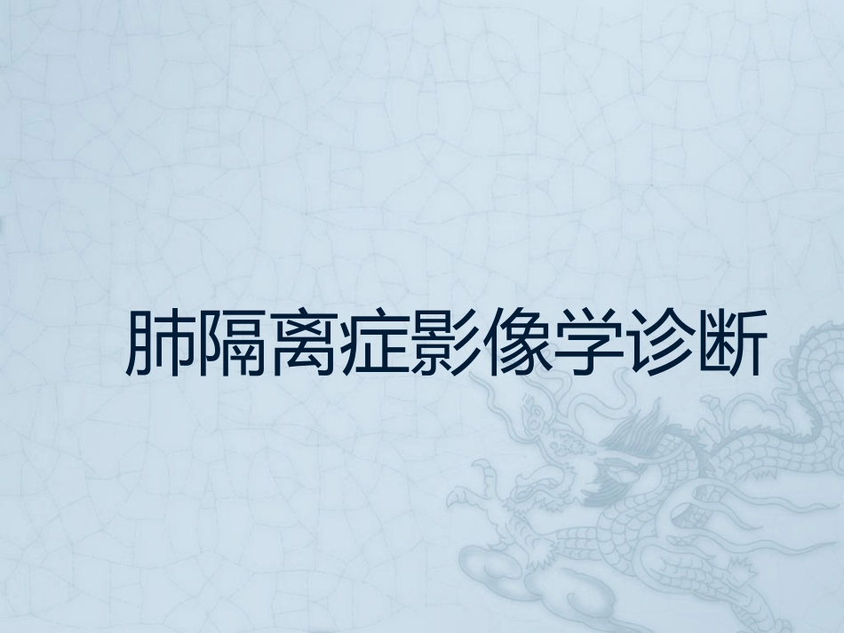
肺隔离症影像学诊断A48-year-oldKoreanfemalepatientpresentedwithanabnormalmasslesionthatwasdetectedbyabdominalcomputedtomographyinavisittoourhospital
Shehadexperiencedintermittentabdominalpainforseveralmonths
Shehadnootherspecificpastmedicalhistoryandnohistoryoftrauma
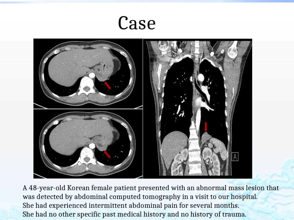
Case10/23/202409:42AM3男性,46岁
反复咳嗽、咳痰多年
胸片及CT,诊断为左下肺支扩并感染、左下肺化脓性病变等
(A)Incisionofdiaphragm(blackarrow),Intradiaphragmaticmass(redarrow)wasidentified
(B)Diaphragmaticbulge(blackarrow)
Incisionsiteofdiaphragm(redarrow)
(C)Smallaberrantvesselswereclipped(blackarrow)
(D)Yellowishmucoidmaterialsweredrained(blackarrow)(A)Grossfindings
(B)Dilatedmucin-filledairwaysandremnantsofcartilaginousbronchi(x100,hematoxylinandeosinstain)
(C)Normallungtissueisnotobserved(x100,hematoxylinandeosinstain)
(D)Dilatedairwaysarelinedbybronchiolartypeepithelium(