中国修复重建外科杂志2008年2月第22卷第2期·145·组织工程骨膜异体体内成骨修复兔骨缺损的初步观察赵琳1史志勇2周晟3贾有福3刘佳1王军胜1【摘要】目的探讨以兔BMSCs和猪SIS复合构建的组织工程骨膜,在异体兔体内成骨的可行性。方法取新西兰大白兔BMSCs与SIS复合构建组织工程骨膜。选2月龄新西兰大白兔12只,制备双侧桡骨1.5~2.0cm缺损模型。随机选一侧植入组织工程骨膜,作为实验组;另一侧仅植入单纯SIS,作为对照组。术后观察动物一般情况,4周后摄X线片观察,取骨缺损中段标本行HE及Masson染色观察。结果两组动物术后饮食及日常活动基本正常;伤口无红肿、溢脓等;伤肢基本能负重行走。
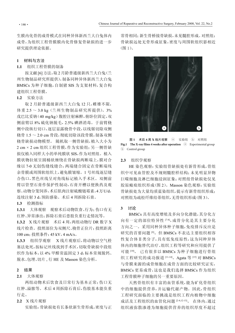
X线片观察:实验组骨缺损处有长条状新生骨形成,密度与正常骨相同,新生骨桥接骨缺损;对照组骨缺损无骨形成征象,骨缺损处密度与周围软组织影相近。
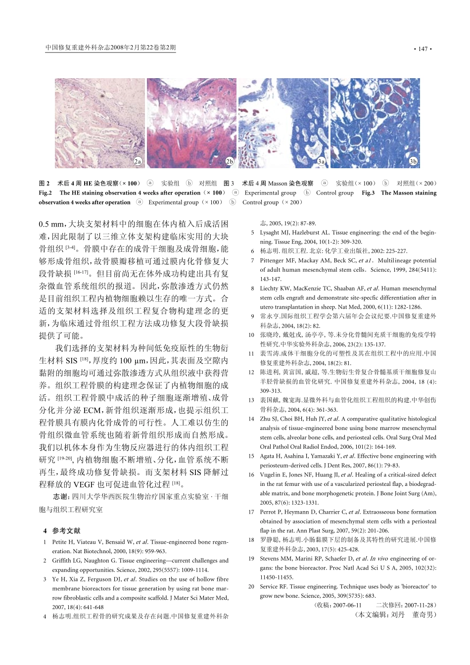
组织学观察:实验组骨缺损处有新骨形成,骨组织中可见血管腔及不规则髓腔样结构;未见明显异物巨噬细胞及淋巴细胞浸润征象;对照组骨缺损处仅为胶原瘢痕组织,无骨组织形成。结论以SIS和BMSCs构建的组织工程骨膜在同种异体体内可以成骨,有修复骨缺损的可行性。【关键词】组织工程骨膜BMSCsSIS骨缺损兔中图分类号:R318.08Q813文献标志码:ATHEPRIMARYOBSERVATIONOFTISSUEENGINEEREDPERIOSTEUMOSTEOGENESISINVIVOINALLO-GENICRABBIT/ZHAOLin1,SHIZhiyong2,ZHouShen3,JIAYoufu3,LIUJia1,WANGJunsheng1.1DepartmentofOrthope-dics,the2ndhospitalofLanzhouuniversity,LanzhouGansu,730030,P.R.China;2TheExperimentalAnimalCenterofGeneralHospitalofLanzhouMilitaryRegion;3DepartmentofRadiology,ChinesetraditionalMedicinehospitalofGansuProvince.Cor-respondingauthor:ZHAOLin,E-mail:bonezl@sina.com【Abstract】ObjectiveToinvestigatetheinvivoosteogenicfeasibilityoftissueengineeredperiosteumconstructedbyporcineSISandBMSCsinallogenicNewZealandrabbit.MethodsThetissueengineeredperiosteumconstructedbySISscaffoldandBMSCswaspreparedinvitro.Twelve2-month-oldNewZealandrabbitswereusedintheexperiments.The1.5-2.0cmcriticalbonedefectsweremadeinthebothsidesofradiusoftheanimals.Thetissueengineeredperiosteumwasgraftedintoonesidedefectrandomly,whiletheothersidedefectwasonlygraftedSIS.Fourweeksafteroperation,theforearmsofallanimalswerecheckedbyX-ray.Then,animalsweresacrificedtoharvestthespecimenwhichweretreatedpromptlyforHEandMassonstaining.TheX-rayfilmandthemorphologicaltissuestainingoutcomewereevaluatedqualitatively.ResultsAfteroperation,allanimalshadanormalbehavioranddiet;theincisionhealednormally;theforearmcouldmovenormallyforbearingweight.ThetissueengineeredperiosteumconstructedbyallogenicBMSCsandheterogeneicSISscaffoldcouldformnewbonetissue,andbridgedthebonedefectwhichcouldbeconfirmedeitherinX-rayfilmorhistologicalstaining.Thenewlyformedbonetissuehadsimilarbonedensitytonormalbone.Alotofirregularnewlyformedvesselsandmedullarycavityinsertedinthenewlybornedtissue.Nolymphocytesinfiltratedinhistologicalexamination.WhilethecontrolsidehadnoanyosteogenesisneithterinX-ray,norinHEandMassonstaininginspecting;thedefectspaceonlyoccupiedwithsomeconnectivetissue.ConclusionTissueengineeredperiosteumcanformnewboneinallogenicrabbitandhasthefeasibilitytorepairthesegmentaldiaphysisdefect.【Keywords】TissueengineeredperiosteumBMSCsSISBonedefectRabbitsFoundationitem:NationalNaturalScienceFoundationofChina(30500133)骨组织的形成主要有膜内化骨及软骨内化骨两种形式,其中膜内化骨在骨折愈合、骨缺损修复的新骨形成中起重要作用。自从Lang等(1989)报道了采用组织工程方法再生骨组织以来,骨缺损修复这一临床难题找到了新的解决途径。虽然骨组织工程的研究很多,基金项目:国家自然科学基金资助项目(30500133)...