实用医学影像杂志2009年第10卷第6期JPMI,2009,Vol.10,No.6卵巢原发肿瘤有50%~70%来源于卵巢体腔上皮,其中以卵巢囊腺瘤与囊腺癌最常见。笔者搜集了32例43个卵巢囊腺肿瘤的CT影像资料作回顾性分析。1资料与方法搜集本院2000年至2007年卵巢肿瘤患者32例,共43个肿瘤,发病年龄18~83岁,平均56岁。所有病例均手术病理证实。临床主要症状有腹痛、腹胀、盆腔包块和绝经后阴道流血等。10例无症状,为体检时B超发现。患者于扫描前4h分4次口服2%的含碘溶液1500mL,待膀胱充盈满意后采用PHILIPS螺旋CT/Aura行全腹平扫和增强扫描,螺距1.0,层厚7mm。造影剂为碘海醇[碘的质量浓度:P(I)=300g/L],注射速率为2.5mL/s,延时60s扫描。卵巢肿瘤可分3型。Ⅰ型为囊性;Ⅱ型为囊实型,又分为3个亚型:Ⅱa型以囊性为主(囊性成分大于2/3),Ⅱb型混合型(实性成分占1/3~2/3),Ⅱc型以实性为主(实性成分大于2/3);Ⅲ型为实性。肿瘤的壁与囊内间隔厚薄以3mm为界,并有规则与否之分;Ⅰ、Ⅱa型有单房或多房之分;Ⅱ型部分可出现囊内壁结节或不规则软组织成分;所有类型均可出现囊外壁结节[1,2]。2结果本组卵巢囊腺瘤:共10例11个肿瘤,CT发现全部病灶。
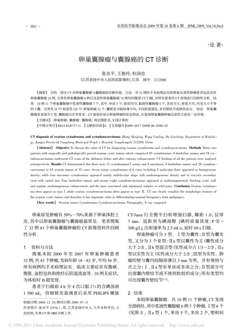
其中浆液性囊腺瘤6例7个肿瘤,Ⅰ型6个(见图1),Ⅱa型1个,单房5个,多房2个,壁和间卵巢囊腺瘤与囊腺癌的CT诊断张水平,王艳玲,杜国忠(江苏省扬中市人民医院影像科,江苏扬中212200)【摘要】目的探讨CT对卵巢囊腺瘤与囊腺癌的诊断价值。方法对32例经手术病理证实的卵巢良恶性肿瘤患者包括良性卵巢囊腺瘤10例,交界性卵巢囊腺瘤4例以及恶性卵巢囊腺癌18例均经腹部CT扫描,对所有患者的CT表现进行回顾性分析。结果10例11个卵巢囊腺瘤中浆液性囊腺瘤7个,其中,单房5个,密度均匀;黏液性囊腺瘤4个,多房为主,密度不均,内见大小不等的子囊。交界性(4个)和恶性(28个)卵巢肿瘤32个,囊壁及分隔厚薄不均,不同程度强化,多有壁结节或肿块成分。结论卵巢囊腺瘤常表现为Ⅰ型,囊腺癌以Ⅱ型常见。CT能很好显示卵巢肿瘤的形态特征,在鉴别卵巢囊腺肿瘤良恶性方面有一定价值。【关键词】卵巢肿瘤;囊腺瘤;囊腺癌;体层摄影术,X线计算机【中图分类号】R814.42;R737.31【文献标识码】A【文章编号】1009-6817(2009)06-0380-03CTdiagnosisofovariancystadenomaandcystadenocarcinomaZhangShuiping,WangYanling,DuGuozhong.DepartmentofRadiolo-gy,JiangsuProvincialYangzhongMunicipalPeople'sHospital,Yangzhongshi212200,Chian【Abstract】ObjectiveTodiscussthevalueofCTfordiagnosingovariancystadenomaandcystadenocarcinoma.MethodsThirty-twopatientswithsurgicallyandpathologicallyprovedovariancystictumorswhichcomprised10cystadenomas,4borderlinetumorsand18cys-tadenocarcinomasunderwentCTscansoftheabdomenbeforeandaftercontrast-enhancement.CTfindingsofallthepatientswereanalyzedretrospectively.ResultsCTdemonstratedthattherewere11cystadenomas(7serousand4mucinous),4borderlinetumorsand28cystadeno-carcinomasin43ovariantumorsof32cases.Sevenserouscystadenomasof6casesincluding5unilocularthoseappearedashomogeneousdensity,whilefourmucinouscystadenomasappearedmainlymultilocularshapewithnonhomogeneousdensityandintracysticsecondarycystswithvariedsize.Fourborderlinetumorsandtwenty-eightcystadenocarcinomasappearedasnonhomogeneouslythickingcysticwallandseptum,nonhomogeneousenhancement,andthemostassociatedwithintramuralnodulesorsolidparts.ConclusionOvariancystadeno-maoftenappearastypeⅠ,whileovariancystadenocarcinomaoftenappearastypeⅡ.CTcanclearlyvisualizethemorphologicfeaturesoftheovariancystictumorsandthereforeithasimportantvalueindifferentiatingtumoralbenignancyfrommalignancy.【Keywords】Ovariantumor;Cystadenoma;Cystadenocarcinoma;Tomography,X-raycomputed收稿日期:2008-12-26;修回日期:2009-07-31作者简介:张水平(1976-),男,江苏...