电脑桌面
添加小米粒文库到电脑桌面
安装后可以在桌面快捷访问

海绵状血管瘤怎么治

海绵状血管瘤怎么治_第1页
1/4
海绵状血管瘤怎么治_第2页
2/4
海绵状血管瘤怎么治_第3页
3/4
下载后可任意编辑【海绵状血管瘤铜针埋置术后的病理分析】海绵状血管瘤怎么治【海绵状血管瘤铜针埋置术后的病理分析】海绵状血管瘤怎么治 【摘要】 目的 分析铜针治疗血管瘤后的组织病理学改变,评价其疗效以及相关影响,探究其方法的改进。方法 收集铜针埋置治疗后 1~13 年的血管瘤标本 13 例,采纳 HE 染色方法分析其病理学改变。
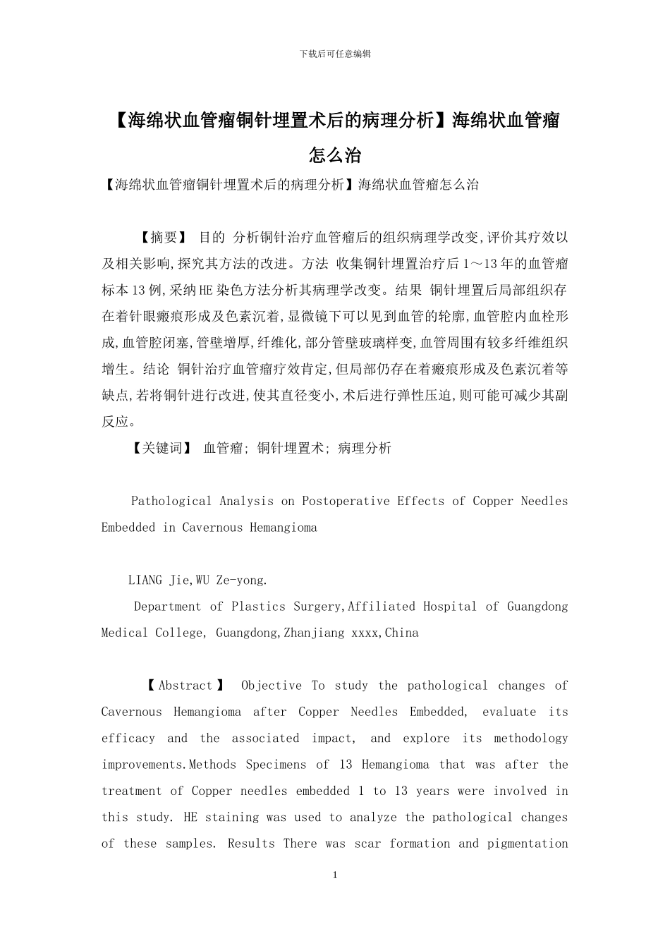
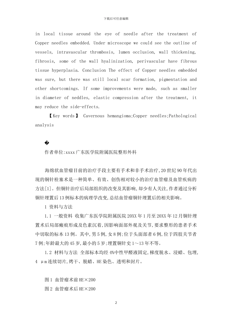
结果 铜针埋置后局部组织存在着针眼瘢痕形成及色素沉着,显微镜下可以见到血管的轮廓,血管腔内血栓形成,血管腔闭塞,管壁增厚,纤维化,部分管壁玻璃样变,血管周围有较多纤维组织增生。结论 铜针治疗血管瘤疗效肯定,但局部仍存在着瘢痕形成及色素沉着等缺点,若将铜针进行改进,使其直径变小,术后进行弹性压迫,则可能可减少其副反应。 【关键词】 血管瘤; 铜针埋置术; 病理分析 Pathological Analysis on Postoperative Effects of Copper Needles Embedded in Cavernous Hemangioma LIANG Jie,WU Ze-yong. Department of Plastics Surgery,Affiliated Hospital of Guangdong Medical College, Guangdong,Zhanjiang xxxx,China 【 Abstract 】 Objective To study the pathological changes of Cavernous Hemangioma after Copper Needles Embedded, evaluate its efficacy and the associated impact, and explore its methodology improvements.Methods Specimens of 13 Hemangioma that was after the treatment of Copper needles embedded 1 to 13 years were involved in this study. HE staining was used to analyze the pathological changes of these samples. Results There was scar formation and pigmentation 1下载后可任意编辑in local tissue around the eye of needle after the treatment of Copper needles embedded. Under microscope we could see the outline of vessels, intravascular thrombosis, lumen occlusion, wall thickening, fibrosis, some of the wall hyalinization, perivascular have fibrous tissue hyperplasia. Conclusion The effect of Copper needles embedded was sure, but there was still local scar formation, pigmentation and other shortcomings. If some improvements were ma...

1、当您付费下载文档后,您只拥有了使用权限,并不意味着购买了版权,文档只能用于自身使用,不得用于其他商业用途(如 [转卖]进行直接盈利或[编辑后售卖]进行间接盈利)。
2、本站所有内容均由合作方或网友上传,本站不对文档的完整性、权威性及其观点立场正确性做任何保证或承诺!文档内容仅供研究参考,付费前请自行鉴别。
3、如文档内容存在违规,或者侵犯商业秘密、侵犯著作权等,请点击“违规举报”。

碎片内容

海绵状血管瘤怎么治

确认删除?
VIP
微信客服
  • 扫码咨询
会员Q群
  • 会员专属群点击这里加入QQ群
客服邮箱
回到顶部