Mimics及脊柱模型用于下颈椎椎弓根个体化置钉的应用研究王远政1,田晓滨1,刘洋2,李波1,孙立1,张一1,王楠筑1(550002贵阳,贵州省人民医院骨科1;400010重庆,重庆医科大学附属第二医院骨科2)[摘要]目的利用快速成型技术及Mimics软件设计一种新的下颈椎椎弓根钉个体化置入技术,并探讨其临床应用意义。方法对16例成人下颈椎标本行CT扫描收集数据,导入Mimics软件对标本进行三维重建。
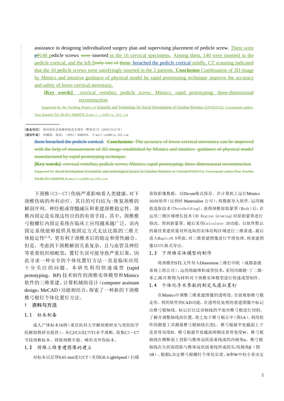
利用Mimics相关功能在三维重建图像上寻找下颈椎椎弓根最佳轴线并测量椎弓根相关参数,制定椎弓根螺钉个体化置入方案。然后将三维重建图像以STL格式导入三维打印机,制作出下颈椎的实体模型,根据个体化置钉角度置入导向针。
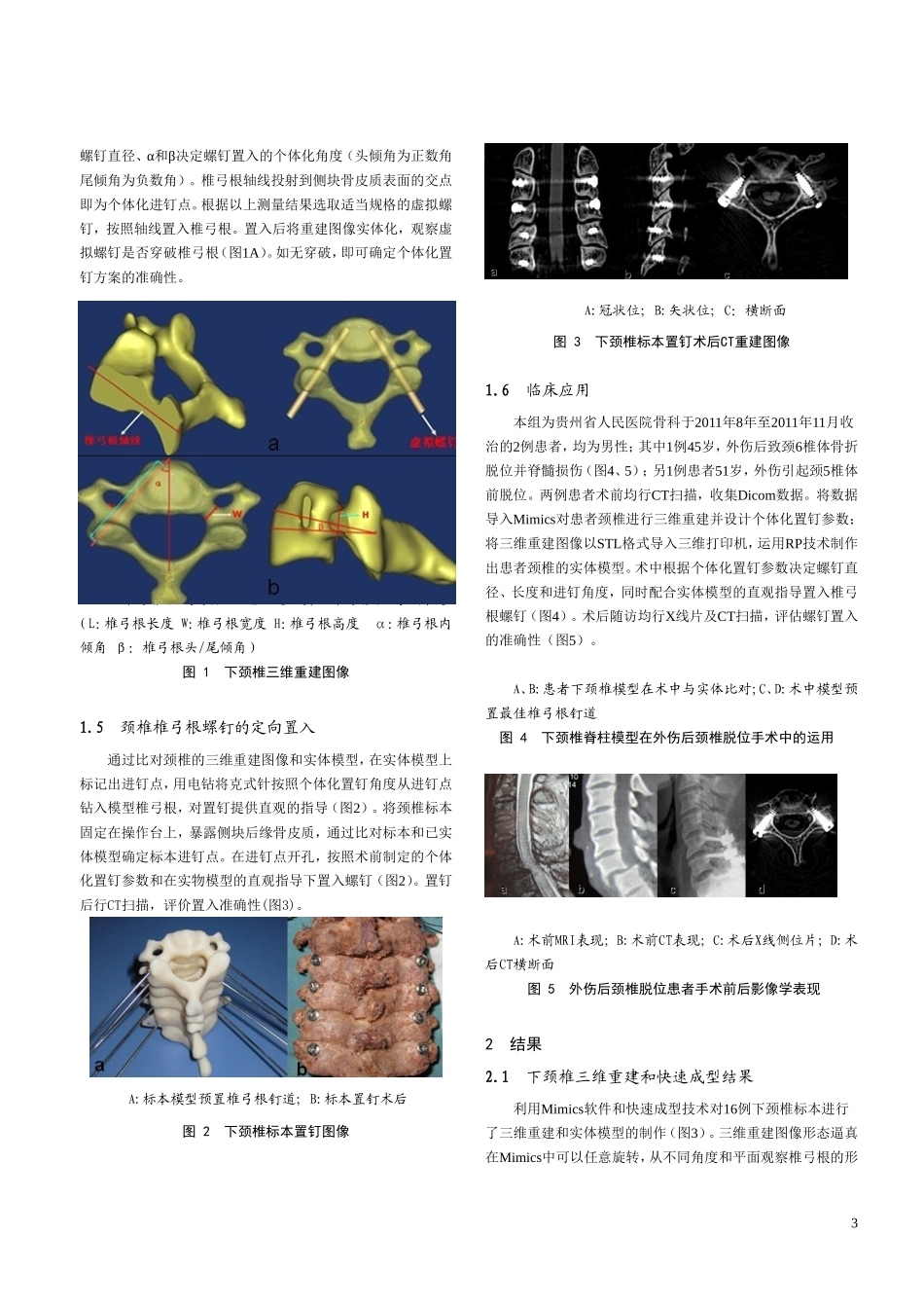
依照制定的个体化指定参数,并配合实体模型的直观指导,在标本上进行置钉。置钉后标本行CT扫描,判断置入准确性。利用上述方法对2例患者进行个体化置钉,术后通过CT扫描验证螺钉位置准确性。结果成功建立了与标本相似度极高的下颈椎三维重建图像和实物模型,通过测量结果设计了每个椎弓根的置钉参数。共在标本上置入148枚椎弓根螺钉,140枚位于椎弓根骨皮质之内,8枚稍穿破椎弓根骨皮质。对患者置入10枚椎弓根螺钉,CT示螺钉位置满意。结论用Mimics软件对下颈椎进行三维重建,制定个体化置钉参数,同时配合实物模型的直观指导,提供了一种下颈椎椎弓根钉个体化置钉的方法,利用该法能提高置钉安全性。[关键词]颈椎;椎弓根螺钉;Mimics软件;快速成型;三维重建[中图法分类号][文献标志码]AIndividualizationoflowercervicalpediclescrewfixationwithApplicationofrapidprototypingandMIMICSimicssoftwareinlowercervicalpediclescrewfixationthree-dimensional(3D)reconstructionmodelwasgeneratedwithMIMICSaworkstationrunningMIMICS10.01softwareWangYuanzheng1,TianXiaobin1,LiuYang2,LiBo1,SunLi1,ZhangYi1,WangNanzhu1(1DepartmentofOrthopaedicsSurgery,GuizZhouProvinceialPeople’sHospital,Guiyang,GuizhouProvince,550002,China;2DepartmentofOrthopaedicsSurgery,XinqiaoSecondaffiliatedHospital,ChongqingMedicalUniversity,Chongqing,400010,China)[Abstract]ObjectiveTodesignanewlowercervicalpediclescrewplacementbasedonstudytheapplicationofMimicssoftwareandrapidprototypingtechnologyandevaluateitsclinicalvalueinlowercervicalpediclescrewplacement,andexploreitsclinicapplication.MethodsCTscansscanningwasperformedofon16adultcadavericcervicalspecimens(C3to-C7).wereperformed.TheobtainedCTdatawereimportedintoaworkstationrunningMIMICSreverseengineeringsoftwareMimicstogenerateestablishcervicalthreedimensional(3D)reconstructionimageswhichsavedasSTLfiles.ThentheseSTLfileswereusedtomanufactureproducecervicalphysicalmodelsusingrapidprototypingtechnique.Theoptimaltrajectorypediclewasexploredandmarkedon3DimagesusingcomputerassistantaideddesignmoduleofMimics,andtherelatedparametersofcervicalpedicleweremeasuredusingmeasurementtoolsofMimics.Sotheindividualizedsurgeryplanofpediclescrewfixationwasdeterminedaccordingtothelocationofpedicletrajectoryandparameters.Thetrajectorypinsweredrilledintophysicalmodeltosupervisetheentrypointandorientationofpediclescrew.Withthevisualizedguidanceofphysicalmodel,pediclescrewswereinsertedincadavericspecimensinstrictaccordancewithindividualizedsurgeryplandeterminedpreviously.Pediclescrewfixationwasperformedintwo2patientswithcervicaldisordersrequiringinstrumentationusingthisindividualizedsurgerymethodasmentionedabove.ResultsThecervical3Dimageandphysicalmodelwerefoundtobeanatomicallysimilarwitheachotherandofgreat1assistanceindesigningindividualizedsurgeryplanandsupervisingplacementofpediclescrew.TherewerepP148pediclescrewswereinsertedinthe16cervicalspecimens,.Amongthem,140wereinsertedinthepedi...