CHINESEJOURNALOFCTANDMRI,FEB2010,Vol.8,No.1totalNo.30·25通讯作者:(200011)上海市第二人民医院放射科裴林惠先天性支气管囊肿的影像诊断及病理学分析上海市第二人民医院放射科裴林惠复旦大学附属中山医院放射科丁建国【摘要】目的先天性支气管囊肿的影像诊断及病理特点。方法回顾性分析我院9例经手术病理证实的先天性支气管囊肿的影像诊断表现。结果根据X线胸片、胸部CT检查结果,本组患者病灶肺内型5例,纵隔型3例,异位型1例。其中液囊肿4例、气囊肿3例、多发肺囊肿2例。
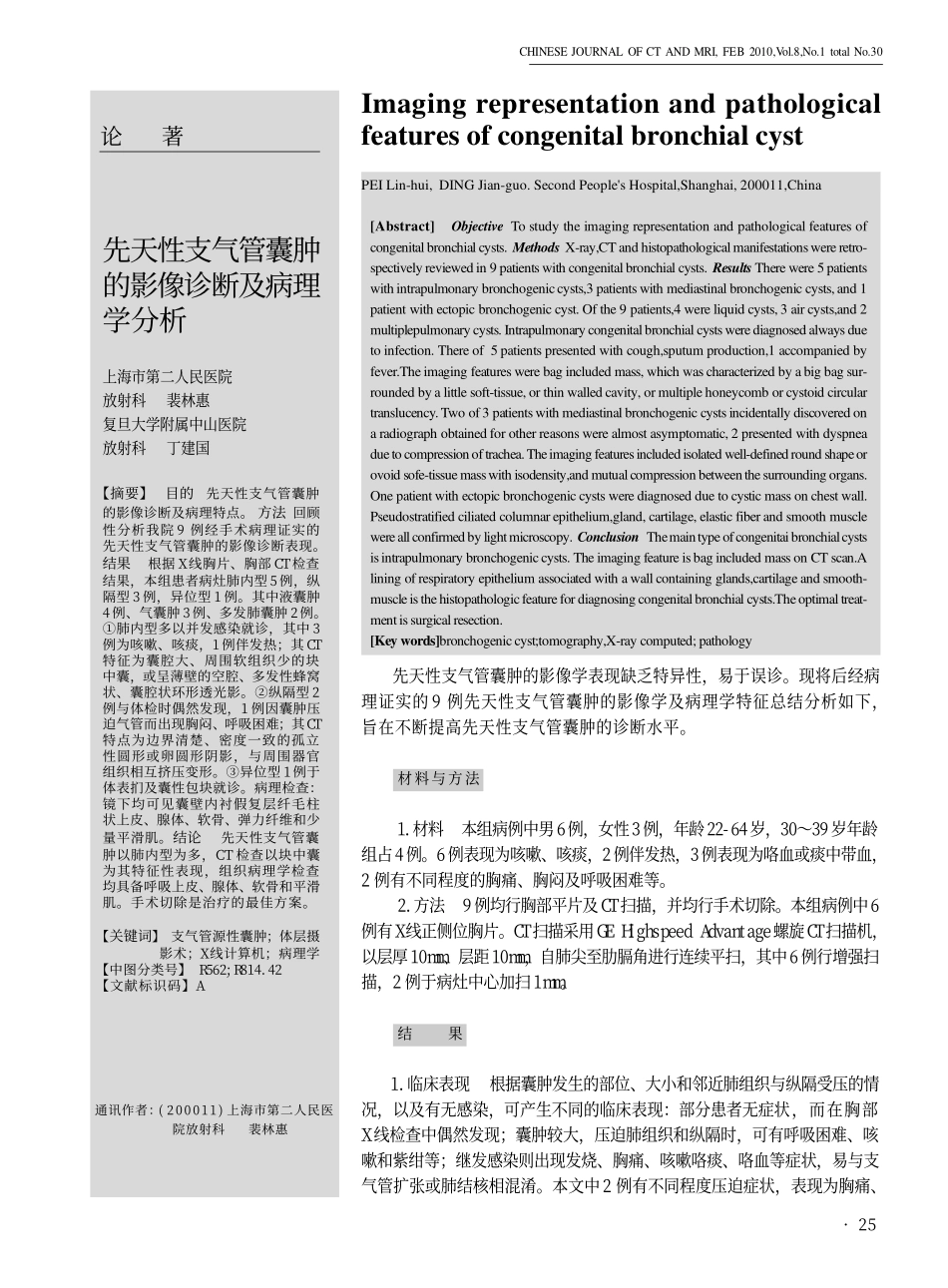
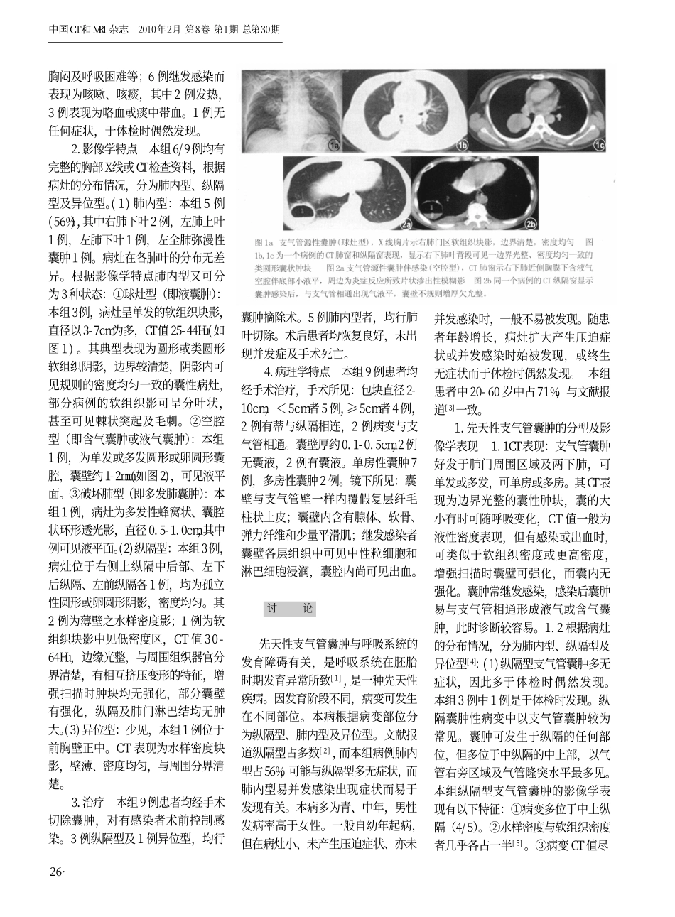
①肺内型多以并发感染就诊,其中3例为咳嗽、咳痰,1例伴发热;其CT特征为囊腔大、周围软组织少的块中囊,或呈薄壁的空腔、多发性蜂窝状、囊腔状环形透光影。②纵隔型2例与体检时偶然发现,1例因囊肿压迫气管而出现胸闷、呼吸困难;其CT特点为边界清楚、密度一致的孤立性圆形或卵圆形阴影,与周围器官组织相互挤压变形。③异位型1例于体表扪及囊性包块就诊。病理检查:镜下均可见囊壁内衬假复层纤毛柱状上皮、腺体、软骨、弹力纤维和少量平滑肌。结论先天性支气管囊肿以肺内型为多,CT检查以块中囊为其特征性表现,组织病理学检查均具备呼吸上皮、腺体、软骨和平滑肌。手术切除是治疗的最佳方案。【关键词】支气管源性囊肿;体层摄影术;X线计算机;病理学【中图分类号】R562;R814.42【文献标识码】AImagingrepresentationandpathologicalfeaturesofcongenitalbronchialcystPEILin-hui,DINGJian-guo.SecondPeople'sHospital,Shanghai,200011,China[Abstract]ObjectiveTostudytheimagingrepresentationandpathologicalfeaturesofcongenitalbronchialcysts.MethodsX-ray,CTandhistopathologicalmanifestationswereretro-spectivelyreviewedin9patientswithcongenitalbronchialcysts.ResultsTherewere5patientswithintrapulmonarybronchogeniccysts,3patientswithmediastinalbronchogeniccysts,and1patientwithectopicbronchogeniccyst.Ofthe9patients,4wereliquidcysts,3aircysts,and2multiplepulmonarycysts.Intrapulmonarycongenitalbronchialcystswerediagnosedalwaysduetoinfection.Thereof5patientspresentedwithcough,sputumproduction,1accompaniedbyfever.Theimagingfeatureswerebagincludedmass,whichwascharacterizedbyabigbagsur-roundedbyalittlesoft-tissue,orthinwalledcavity,ormultiplehoneycomborcystoidcirculartranslucency.Twoof3patientswithmediastinalbronchogeniccystsincidentallydiscoveredonaradiographobtainedforotherreasonswerealmostasymptomatic,2presentedwithdyspneaduetocompressionoftrachea.Theimagingfeaturesincludedisolatedwell-definedroundshapeorovoidsofe-tissuemasswithisodensity,andmutualcompressionbetweenthesurroundingorgans.Onepatientwithectopicbronchogeniccystswerediagnosedduetocysticmassonchestwall.Pseudostratifiedciliatedcolumnarepithelium,gland,cartilage,elasticfiberandsmoothmusclewereallconfirmedbylightmicroscopy.ConclusionThemaintypeofcongenitaibronchialcystsisintrapulmonarybronchogeniccysts.TheimagingfeatureisbagincludedmassonCTscan.Aliningofrespiratoryepitheliumassociatedwithawallcontainingglands,cartilageandsmooth-muscleisthehistopathologicfeaturefordiagnosingcongenitalbronchialcysts.Theoptimaltreat-mentissurgicalresection.[Keywords]bronchogeniccyst;tomography,X-raycomputed;pathology论著先天性支气管囊肿的影像学表现缺乏特异性,易于误诊。现将后经病理证实的9例先天性支气管囊肿的影像学及病理学特征总结分析如下,旨在不断提高先天性支气管囊肿的诊断水平。材料与方法1.材料本组病例中男6例,女性3例,年龄22-64岁,30~39岁年龄组占4例。6例表现为咳嗽、咳痰,2例伴发热,3例表现为咯血或痰中带血,2例有不同程度的胸痛、胸闷及呼吸困难等。2.方法9例均行胸部平片及CT扫描,并均行手术切除。本组病例中6例有X线正侧位胸片。CT扫描采用GEHighspeedAdvantage螺旋CT扫描机,以层厚10m...