肺脓肿影像诊断Theimagingdiagnosisoflungabscess24/10/241主要内容1
肺脓肿临床及病理3
肺脓肿影像诊断4
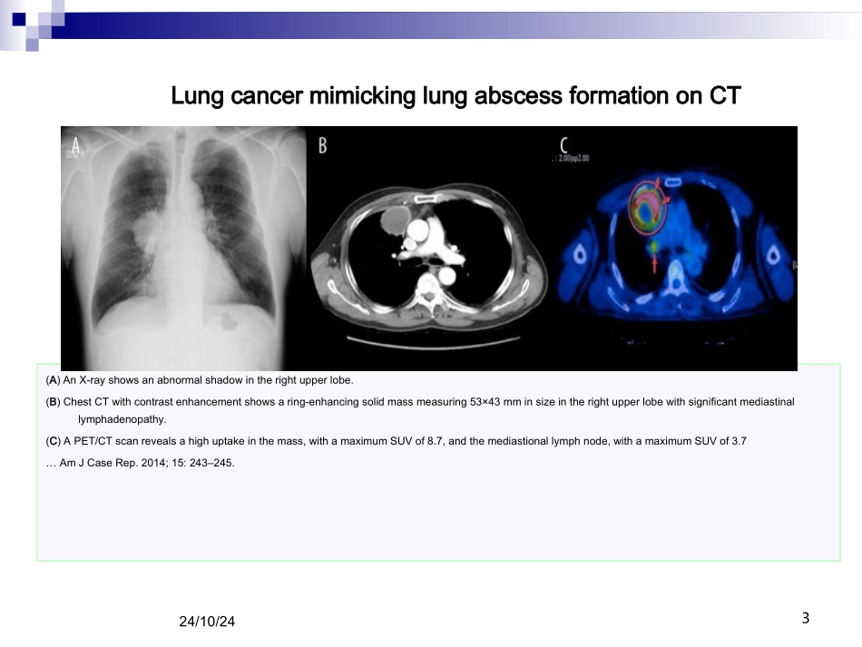
小结(A)AnX-rayshowsanabnormalshadowintherightupperlobe
(B)ChestCTwithcontrastenhancementshowsaring-enhancingsolidmassmeasuring53×43mminsizeintherightupperlobewithsignificantmediastinallymphadenopathy
(C)APET/CTscanrevealsahighuptakeinthemass,withamaximumSUVof8
7,andthemediastionallymphnode,withamaximumSUVof3
7…AmJCaseRep
2014;15:243–245
LungcancermimickinglungabscessformationonCT24/10/243Afollow-upCTscanshowsanincreaseinthesizeofthemass59×49mm(53×43mm)Thefinalresultsofthepathologicexaminationshowedapleomorphiccarcinomafollow-up24/10/244Figure1
Chestradiographshowsalargecavitywithairfluidlevelinleftlung
Figure2
CECTThoraxatthelevelofcarinashowingalargecavitywithairfluidlevelinleftlung
Wallofthecavityisirregul