电脑桌面
添加小米粒文库到电脑桌面
安装后可以在桌面快捷访问

前列腺不典型小腺泡增生的病理形态及临床意义VIP免费

前列腺不典型小腺泡增生的病理形态及临床意义_第1页
1/4
前列腺不典型小腺泡增生的病理形态及临床意义_第2页
2/4
前列腺不典型小腺泡增生的病理形态及临床意义_第3页
3/4
�论著�作者单位:100853北京,解放军总医院病理科通信作者:韦立新(Emai:lweilx301@263.net)前列腺不典型小腺泡增生的病理形态及临床意义石怀银�韦立新�周振鸿�文载律��摘要!�目的�探讨前列腺不典型小腺泡增生的形态学特点和临床意义。方法�收集解放军总医院病理科2004∀2006年前列腺穿刺活检诊断为不典型小腺泡增生病例11例,复习HE和免疫组织化学切片,并对有不典型小腺泡增生病变的蜡块重新进行34�E12、p63和P504S免疫组织化学(SP法)染色,观察不典型小腺泡增生的组织学特点和免疫组织化学表达特点。
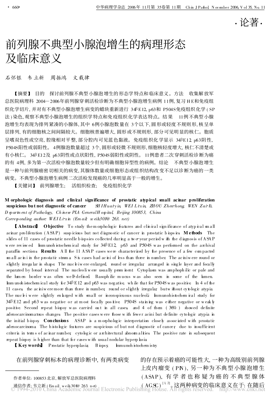
结果�11例不典型小腺泡增生均表现为排列紧凑的小腺体,其中6例小腺泡数量在3个以下,圆形或轻度不规则形,核呈单层排列,有的细胞核之间间隔较大。细胞核普遍增大,圆形或不规则形,部分可见明显的核仁。胞质呈嗜双色性或空亮,腔缘相对平整,部分腔内可见蓝色黏液。免疫组织化学显示34�E12、p63阴性,P504S阳性或弱阳性。4例腺泡数量超过3个,圆形或轻微不规则形,细胞核轻度增大,核仁不清楚或有小核仁。34�E12及p63阴性或点状阳性,P504S弱阳性或阴性。11例患者二次穿刺活检诊断为癌的有4例,多为第一次活检中腺泡数量较少但有明确细胞异型性的病例。结论�不典型小腺泡增生是一种与前列腺癌密切相关的病变,其腺体数量或细胞形态或组织结构改变不足以诊断为癌的一类病变。不典型小腺泡增生病例二次活检发现癌的几率明显高于一般的增生。关键词!�前列腺增生;�活组织检查;�免疫组织化学Morphologicdiagnosisandclinicalsignificanceofprostaticatypicalsmallacinarproliferationsuspiciousbutnotdiagnosticofcancer��SHIHuai�yin,WEILi�xin,ZHOUZhen�hong,WENZai�l�.DepartmentofPathology,ChinesePLAGeneralHospital,Beijing100853,ChinaCorrespondingauthor:WEILi�xin(Email:weilx301@263.net)Abstract!�Objective�Tostudythemorphologicfeaturesandclinicalsignificanceofatypicalsmallacinarproliferation(ASAP)suspiciousbutnotdiagnosticofcancerinprostaticbiopsies.Methods�Theslidesof11casesofprostaticneedlebiopsiescollectedduringatwo�yearperiodwiththediagnosisofASAPwerereviewed.Immunohistochemicalstudyfor34�E12,p63andP504Swasperformedonthearchivalparaffinsections.Results�Allthe11ASAPcaseswerecharacterizedbythepresenceofafewcompactedsmallaciniintheprostaticstroma.Sixcaseshadacinioflessthanthreeinnumber.Theaciniwereroundorslightlyirregularinshape.Thenucleiwereenlarged,roundorirregular,arrangedinsinglelayerandfocallyseparatedbybroadinterva.lThenucleoliwereusuallyprominent.Cytoplasmwasamphophilicorpaleandthelumenborderwasoftenwell�defined.Basophilicmucuswasalsoseeninsomeofthelumen.Immunohistochemicalstudyfor34�E12andp63wasnegative,whilethatforP504Swaspositive.In4ofthe11cases,theaciniweremorethanthreeinnumber,roundorslightlyirregular,butwithoutcytologicatypia.Thenucleiwereslightlyenlargedwithsmallorinconspicuousnucleol.iImmunohistochemicalstudyfor34�E12andp63wasnegativeoratmostfocallypositive.P504Sstainingwaseithernegativeorweaklypositive.Secondrepeatbiopsywascarriedoutinallcases,and4ofthem(36%)showeddefiniteadenocarcinomatouschanges.Thepositivecaseswerethosewithfeweracinibutdefinitecytologicatypiaintheinitialbiopsy.Conclusions�ASAPisamorphologicinterpretationcloselyassociatedwithprostaticadenocarcinoma.Thehistologicfeaturesaresuspiciousofbutnotdiagnosticofcancer,duetoinsufficientcriteriaintermsofacinarnumber,cytologicorarchitecturalabnormalities.Thepositiverateinsubsequentrepeatbiopsyishigherthanthatforcaseswithusualnodularhyperplasia.Keywords!�Prostatichyperplasia;�Biopsy;�Immunohistochemistry��在前列腺穿刺标本的病理诊断中,有两类病变的存在预示着癌的可能性大,一种为高级别前列腺上皮内瘤变(PIN),另...

1、当您付费下载文档后,您只拥有了使用权限,并不意味着购买了版权,文档只能用于自身使用,不得用于其他商业用途(如 [转卖]进行直接盈利或[编辑后售卖]进行间接盈利)。
2、本站所有内容均由合作方或网友上传,本站不对文档的完整性、权威性及其观点立场正确性做任何保证或承诺!文档内容仅供研究参考,付费前请自行鉴别。
3、如文档内容存在违规,或者侵犯商业秘密、侵犯著作权等,请点击“违规举报”。

碎片内容

前列腺不典型小腺泡增生的病理形态及临床意义

确认删除?
VIP
微信客服
  • 扫码咨询
会员Q群
  • 会员专属群点击这里加入QQ群
客服邮箱
回到顶部