原发性肝癌门静脉癌栓MRI和DSA比较朱锡旭陈自谦陈君坤南京军区南京总医院医学影像科(南京,210002)摘要目的:以血管造影表现作为诊断标准,用非侵入性磁共振成像(MRI)检查评价门静脉癌栓。方法:磁共振为1.0T超导系统,轴位扫描序列为SE序列T1权重和T2权重及小角度扫描法(FLASH)序列,数字减影血管造影术(DSA)为PhilipsC2000数字减影血管造影机,造影中采用减影技术。
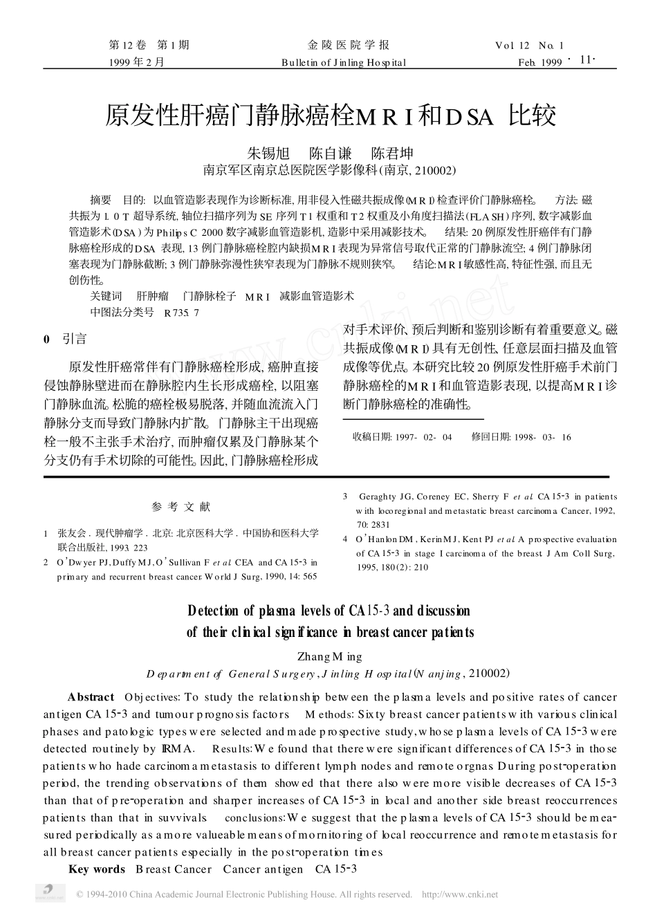
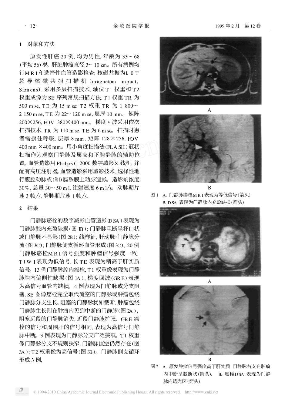
结果:20例原发性肝癌伴有门静脉癌栓形成的DSA表现,13例门静脉癌栓腔内缺损MRI表现为异常信号取代正常的门静脉流空;4例门静脉闭塞表现为门静脉截断;3例门静脉弥漫性狭窄表现为门静脉不规则狭窄。结论:MRI敏感性高,特征性强,而且无创伤性。关键词肝肿瘤门静脉栓子MRI减影血管造影术中图法分类号R735.70引言原发性肝癌常伴有门静脉癌栓形成,癌肿直接侵蚀静脉壁进而在静脉腔内生长形成癌栓,以阻塞门静脉血流。松脆的癌栓极易脱落,并随血流流入门静脉分支而导致门静脉内扩散。门静脉主干出现癌栓一般不主张手术治疗,而肿瘤仅累及门静脉某个分支仍有手术切除的可能性。因此,门静脉癌栓形成对手术评价、预后判断和鉴别诊断有着重要意义。磁共振成像(MRI)具有无创性、任意层面扫描及血管成像等优点。本研究比较20例原发性肝癌手术前门静脉癌栓的MRI和血管造影表现,以提高MRI诊断门静脉癌栓的准确性。收稿日期:1997-02-04修回日期:1998-03-16参考文献1张友会1现代肿瘤学1北京:北京医科大学1中国协和医科大学联合出版社,1993.2232O’DwyerPJ,DuffyMJ,O’SullivanFetal.CEAandCA1523inprimaryandrecurrentbreastcancer.WorldJSurg,1990,14:5653GeraghtyJG,CoreneyEC,SherryFetal.CA1523inpatientswithlocoregionalandmetastaticbreastcarcinoma.Cancer,1992,70:28314O’HanlonDM,KerinMJ,KentPJetal.AprospectiveevaluationofCA1523instageIcarcinomaofthebreast.JAmCollSurg,1995,180(2):210DetectionofplasmalevelsofCA15-3anddiscussionoftheirclinicalsignificanceinbreastcancerpatientsZhangMingDepartmentofGeneralSurgery,JinlingHospital(Nanjing,210002)AbstractObjectives:TostudytherelationshipbetweentheplasmalevelsandpositiveratesofcancerantigenCA1523andtumourprognosisfactors.Methods:Sixtybreastcancerpatientswithvariousclinicalphasesandpatologictypeswereselectedandmadeprospectivestudy,whoseplasmalevelsofCA1523weredetectedroutinelybyIRMA.Results:WefoundthatthereweresignificantdifferencesofCA1523inthosepatientswhohadecarcinomametastasistodifferentlymphnodesandremoteorgnas.Duringpost2operationperiod,thetrendingobservationsofthemshowedthattherealsoweremorevisibledecreasesofCA1523thanthatofpre2operationandsharperincreasesofCA1523inlocalandanothersidebreastreoccurrencespatientsthanthatinsuvvivals.conclusions:WesuggestthattheplasmalevelsofCA1523shouldbemea2suredperiodicallyasamorevalueablemeansofmornitoringoflocalreoccurrenceandremotemetastasisforallbreastcancerpatientsespeciallyinthepost2operationtimes.KeywordsBreastCancerCancerantigenCA1523·11·第12卷第1期金陵医院学报Vol.12No.11999年2月BulletinofJinlingHospitalFeb.19991对象和方法原发性肝癌20例,均为男性,年龄为33~68(平均56)岁。肝脏肿瘤直径3~10cm。所有病例均行MRI和选择性血管造影检查;核磁共振为1.0T超导核磁共振扫描机(magnetomimpact,Siemens),采用多层扫描技术,轴位T1权重和T2权重成像为SE序列常规扫描方法,T1权重TR为500mse,TE为15mse;T2权重TR为1800~2150mse,TE为22~120mse,层厚10mm。矩阵200×256,FOV380×400mm。梯度回波采用依次扫描技术,TR为110mse,TE为6mse。扫描时患者需摒住呼吸,层厚8mm,矩阵128×256,FOV400mm×400mm。用小角度扫描法(FLASH)冠状扫描作为观察门静脉及属支和下腔静脉的辅助位置。血管造影用PhilipsC2000数字减影X线机,并配有高压注射器。血管造影采用减影技术,选择性地行腹腔动脉或(和)肠系膜上动脉造影。造影剂浓度30%,总量30~50ml,注射速度...