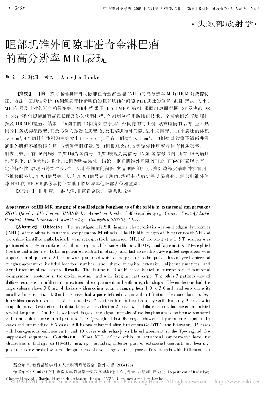
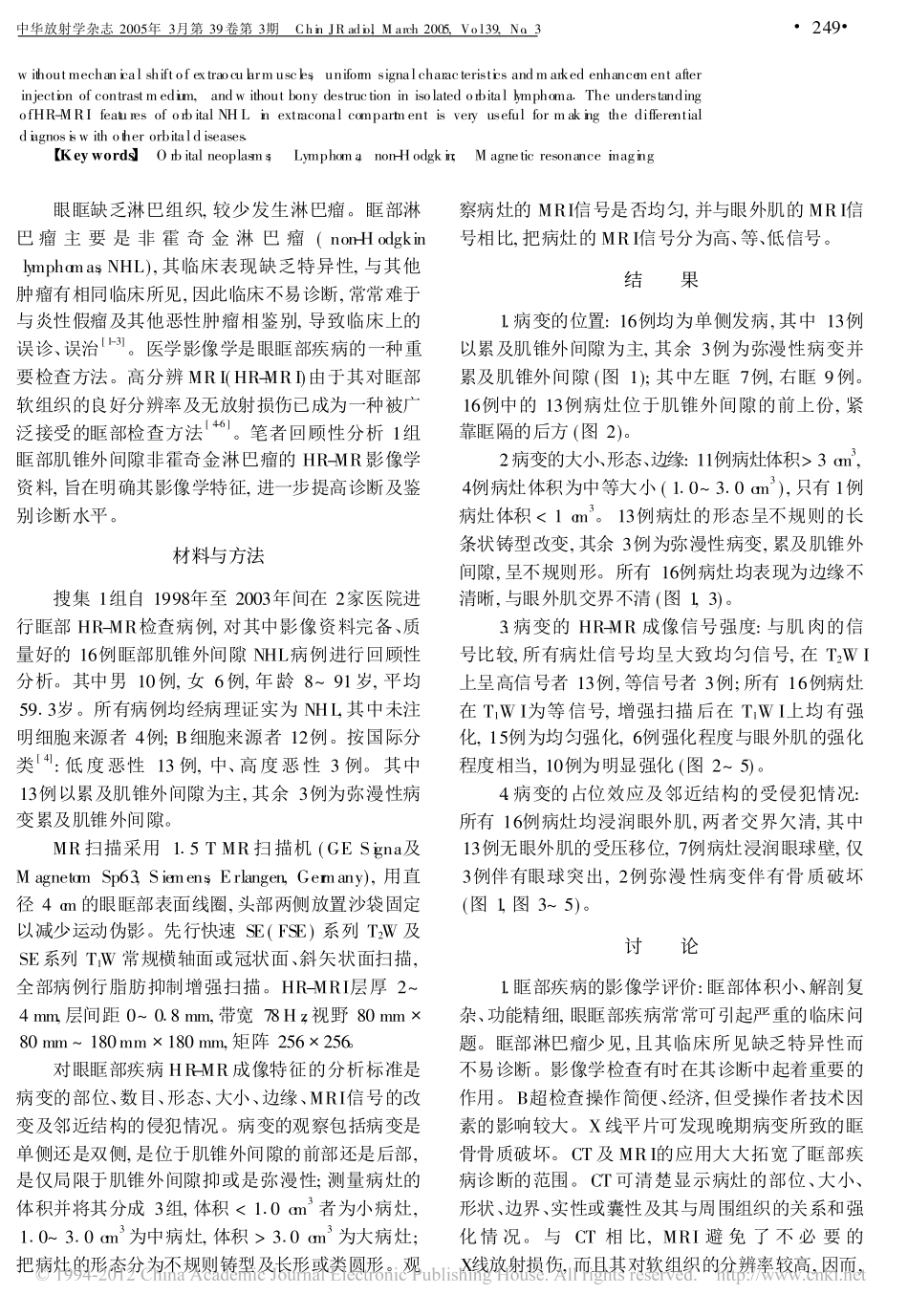
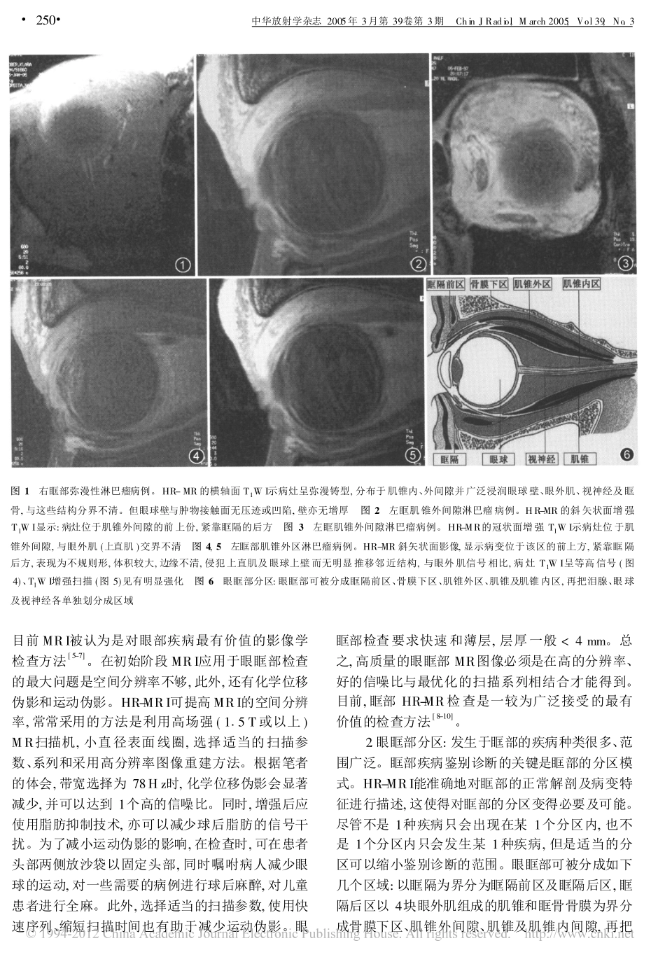
#头颈部放射学#基金项目:教育部留学回国人员科研启动基金(教外司留:2004176)作者单位:510632广州,暨南大学附属第一医院医学影像中心(周全、刘斯润、黄力);DepartmentofRadiology,Virchow-Hospita,lCharitÜ,Humboldt-University,Berlin,13353,Germany(Arne-JrnLemke)眶部肌锥外间隙非霍奇金淋巴瘤的高分辨率MRI表现周全刘斯润黄力Arne-JrnLemke【摘要】目的探讨眶部肌锥外间隙非霍奇金淋巴瘤(NHL)的高分辨率MR(HR-MR)成像特征。方法回顾性分析16例经病理诊断明确的眶部肌锥外间隙NHL病灶的位置、数目、形态、大小、MRI信号及其对邻近结构侵犯等。MR扫描采用115TMR扫描机,眼眶部表面线圈,SE及快速SE(FSE)序列常规横轴面或冠状面及斜矢状面扫描,全部病例行脂肪抑制技术。全部病例均行增强扫描及HR-MRI检查。结果16例中的13例病灶位于肌锥外间隙的前上份,紧靠眶隔的后方,呈不规则的长条状铸型改变;其余3例为弥漫性病变,累及眶部肌锥外间隙,呈不规则形。11个病灶的体积>3cm3,4个病灶的体积为中等大小(1~3cm3),只有1例病灶<1cm3。13例病灶边缘不清晰并浸润眼外肌但不推移眼外肌;7例浸润眼球壁,仅3例眼球突出,2例弥漫性病变者伴有骨质破坏。与肌肉比较,所有16例病灶T1WI均为等信号,T2WI表现为高信号13例,等信号3例;所有16例病灶均有强化,15例为均匀强化,10例为明显强化。结论眶部肌锥外间隙NHL的HR-MRI表现具有一定的特征性,表现为铸型生长、位于肌锥外间隙的前份,紧靠眶隔的后方、病灶边缘欠清晰并浸润,但不推移眼外肌,T1WI信号等于肌肉、T2WI信号高于肌肉、增强扫描病灶呈明显强化。眶部肌锥外间隙NHL的HR-MR影像学特征有助于临床与其他眶部占位相鉴别。【关键词】眶肿瘤;淋巴瘤,非霍奇金氏;磁共振成像AppearanceofHR-MRimagingofnon-HodgkinlymphomasoftheorbitsinextraconalcompartmentZHOUQuan*,LIUSi-run,HUANGLi,Arne-JrnLemke1*MedicalImagingCenter,FirstAffiliatedHospital,JinanUniversityMedicalCollege,Guangzhou510630,China【Abstract】ObjectiveToinvestigateHR-MRimagingcharacteristicsofnon-Hodgkinlymphomas(NHL)oftheorbitsinextraconalcompartment1MethodsTheHR-MRimagesof16patientswithNHLoftheorbitsidentifiedpathologicallywereretrospectivelyanalyzed1MRIoftheorbitata115Tscannerwasperformedwith4-cmsurfacecoi,lthinslice,suitablebandwidth,smallFOV,andlargematrix1T1-weighted(beforeandafteri1v1bolusinjectionofcontrastmedium)andfastspin-echoT2-weightedsequenceswereacquiredinallpatients1Allcaseswereperformedwithfatsuppressiontechniques1Theanalyzedcriteriaofimagingappearanceincludedlocation,number,size,shape,margins,extension,adjacentstructures,andsignalintensityofthelesions1ResultsThelesionsin13of16caseslocatedinanteriorpartofextraconalcompartment,posteriortotheorbitalseptum,andwithirregularcastshape1Theother3patientsshoweddiffuselesionswithinfiltrationinextraconalcompartmentandwithirregularshape1Elevenlesionshadthelargevolumeabove310m,l4lesionswithmediumvolumerangingfrom110to310m,landonlyonewithsmallvolumelessthan110ml113caseshadapoor-definedmarginwithinfiltrationofextraocularmuscles,butwithoutmechanicalshiftofthemuscles17patientshadinfiltrationofeyebal,lbutonly3caseswithexophthalmos1Destructionoforbitalbonewasevidentin2caseswithdiffuselesionsbutneverinisolatedorbitallymphoma1OntheT1-weightedimages,thesignalintensityofthelymphomawasisointensecomparedwiththatofthemuscleinallpatients1TheT2-weightedfastSEimagesshowedahyperintensesignalin13casesandintermediatein3cases1AlllesionsenhancedafterintravenousGd-DTPAadministration,15caseswithhomogeneousenhancement,and10caseswithreliablyvisibleenhancementintheT1-weightedfat-suppressedsequences1ConclusionMostNHLoftheorbitsinextraconalcompartmenthavethecharacteristicfindingsonHR-...