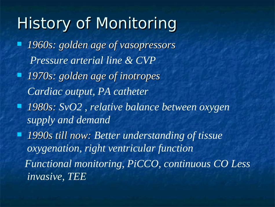
血流动力学监测进展血流动力学监测进展浙江省立同德医院浙江省立同德医院ICUICU陈扬波陈扬波HistoryofMonitoringHistoryofMonitoring1960s:goldenageofvasopressors1960s:goldenageofvasopressorsPressurearterialline&CVP1970s:goldenageofinotropes1970s:goldenageofinotropesCardiacoutput,PAcatheter1980s:1980s:SvO2,relativebalancebetweenoxygensupplyanddemand1990stillnow:1990stillnow:Betterunderstandingoftissueoxygenation,rightventricularfunctionFunctionalmonitoring,PiCCO,continuousCOLessinvasive,TEE血流动力学监测是临床危重病急救的重要内容血流动力学监测是临床危重病急救的重要内容之一,是大手术和抢救危重病员不可缺少的手段。之一,是大手术和抢救危重病员不可缺少的手段。无创伤性血流动力学监测无创伤性血流动力学监测((noninvasivenoninvasivehemodynamicmonitoringhemodynamicmonitoring))创伤性血流动力学监测创伤性血流动力学监测((invasivehemodynamicinvasivehemodynamicmonitoringmonitoring))一、一、无创血流动力学监测(一)心阻抗血流图((一)心阻抗血流图(Impedancecardiogram,ICGImpedancecardiogram,ICG))((二)超声心动图(二)超声心动图(ultrasoniccardiogram,ultrasoniccardiogram,echocardiogram,UCGechocardiogram,UCG))((三)多普勒心排血量监测三)多普勒心排血量监测(四)二氧化碳无创心排血量测定(四)二氧化碳无创心排血量测定(一)心阻抗血流图(一)心阻抗血流图其基本原理是欧姆定律其基本原理是欧姆定律((电阻电阻==电压电压//电流电流))。。1966年Kubicek采用直接式阻抗仪测定心阻抗变化,推导出著名的Kubicek公式。1981年年SramekSramek提出胸腔是锥台型,因此改良了提出胸腔是锥台型,因此改良了KKubicekubicek公式,应用公式,应用88只电极分别安置在颈根部只电极分别安置在颈根部和剑突水平,测量心动周期胸部电阻抗的变化来和剑突水平,测量心动周期胸部电阻抗的变化来测定左心室收缩时间(测定左心室收缩时间(systolictimeinterval,systolictimeinterval,STI)STI)和计算每搏量和计算每搏量,,通过微处理机,自动计算通过微处理机,自动计算CO,CO,并演算出一系列心功能参数。并演算出一系列心功能参数。SV=(Vept·T·△Z/sec)/ZoFigure1:ApplicationofelectrodesinimpedancecardiographyImpedancecardiography(ICG)isasafe,non-invasivemethodtomeasureapatient'shemodynamicstatus.TheICGwaveformisgeneratedbythoracicelectricalbioimpedance(TEB)technology,whichmeasuresthelevelofchangeinimpedanceinthethoracicfluid.Foursmallsensorssendandreceivealowamplitudeelectricalcurrentthroughthethoraxtodetectthelevelofchangeinresistanceinthethoracicfluid.Witheachcardiaccycle,fluidlevelschange,whichaffectstheimpedancetotheelectricalsignaltransmittedbythesensors.ThetechnologybehindICGFigure7:Variationofventricular,aorticandatrialpressure,aorticflow,thoracicimpedancechangeandfistderivativeofimpedance(dz/dt)asafunctionoftime(t).ECGandphonocardiogramtakensimultaneouslyisalsoshown.Thecurvedepictsthecardiacevents/performance.B–OpeningoftheAorticValve,X–ClosureoftheAorticValve,Y–closureofpulmonaryvalve,O–mitralvalveopening/rapidventricularfilling,B-X–VentricularEjectionTime(VET),C–Maximaldeflectionofdz/dt(PeakFlow),B-Cslope–AccelerationContractilityIndex,A–AtrialSystole,Q–StartofventriculardepolarizationPhilipsImpedanceCardiography(ICG)continuouslymeasureshemodynamicparameterswithouttheassociatedrisksoftraditionalinvasivemethods.ThePhilipsICGmeasurementisidealforhemodynamicevaluationofadultpatientsin:EmergencydepartmentsStep-downunitsSpecialprocedureUsingICGfortheappropriatepatientpopulationTheICGmeasurementisdesignedforassessmentofmostadultpat...