·353·http://www.cjmri.cn临床研究|ClinicalArticles磁共振成像2010年第1卷第5期ChinJMagnResonImaging,2010,Vol1,No5[摘要]目的探讨AIDS合并弓形虫脑病的影像学表现及特征。方法收集10例AIDS合并弓形虫脑病的临床及影像学资料,回顾性分析其头颅影像表现特点。
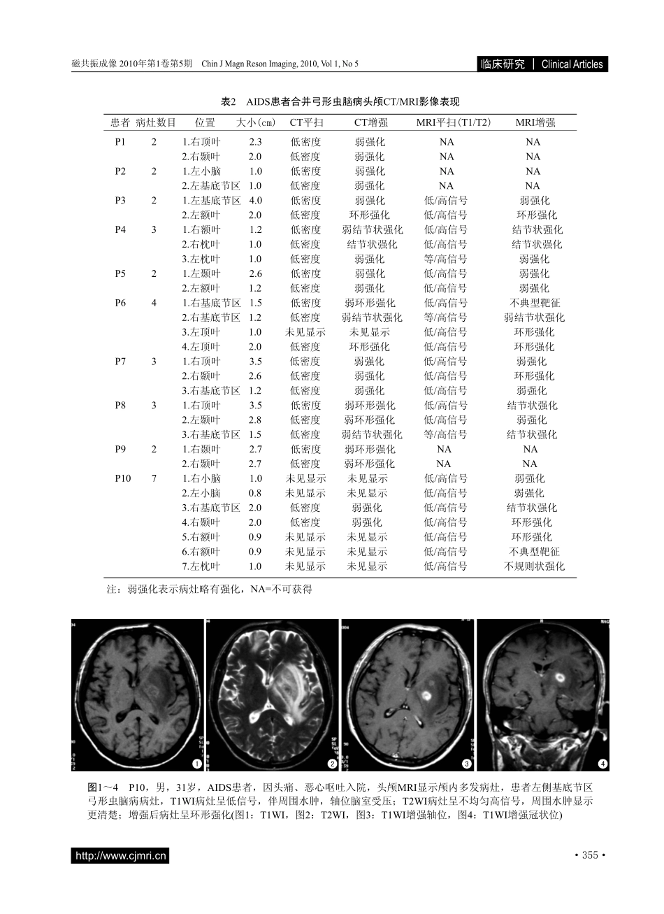
结果10例AIDS合并弓形虫脑病患者中共发现颅内病灶30个,每个患者病灶数目2~7个不等;结节状或类圆形病灶共20个。CT平扫所有颅内病灶表现均为低密度影,增强扫描有7个病灶出现环形强化;MRT1WI表现为低或等信号,T2WI为高信号,增强MRI有8个病灶呈环形强化,其中2个表现为“靶征”,比较CD4+T淋巴细胞计数高于50cell/μl的患者和CD4+T淋巴细胞计数低于50cell/μl的患者,发现前者更易出现环形或结节状强化(卡方检验,P=0.009)。结论AIDS合并弓形虫脑病颅内病灶影像表现为多发性,多累及大脑半球、基底节区及丘脑,病灶的形态多为结节状或类圆形,在CD4+T淋巴细胞计数相对较高的患者注入对比剂后更易呈环形或结节状强化。[关键词]免疫缺陷病毒,人类;获得性免疫缺陷综合征;弓形虫病,脑;计算机断层摄影术;磁共振成像ImagingappearanceoftoxoplasmaencephalopathyinAIDSpatientsHUANGHua,DENGYing-ying,LUPu-xuan*,MAWei,ZHUWen-ke,YANGGen-dong,YERu-xinDepartmentofRadiology,ShenzhenThirdPeople'sHospital,Shenzhen518020,China*Correspondenceto:LuPX,E-mail:lupuxuan@126.comReceived13Jul2010;Accepted25Aug2010AbstractObjective:TocharacterizethecranialimagingfindingsoftoxoplasmaencephalopathyinAIDSpatients.MaterialsandMethods:Aretrospectivereviewof10humanAIDSpatientswithtoxoplasmaencephalopathywasperformed.Headcomputedtomography(CT)andmagneticresonanceimaging(MRI)werereviewed.Results:Therewere30lesionsfoundin10patients,thenumberoflesionsforeachpatientrangedfrom2to7,therewere20nodularorovallesionsamongthe30lesions.Beforecontrastinjection,allthelesionswerelowdensityonCT;afterintravenouscontrastinjection,therewere7lesionsshowedringenhancement.FortheMRI,alllesionswerepredominantlyhypointenseonT1-weightedimages,allthelesionswerehyperintenseonT2-weightedimages.Post-contrastimagingrevealedringenhancementin8lesions,including2targetenhancement.PatientswithCD4+Tlymphocytecountabove50cell/μlcomparedwithCD4+Tlymphocytecountlessthan50cell/μlwasmorelikelywithringornodularenhancement(Chi-squaretest,P=0.009).Conclusion:Toxoplasmaencephalopathyshowedmultiplelesions,manyofthelesionsinvolvedthecerebralhemispheres,basalgangliaandthalamus,thelesionsformwerepredominantlynodularoroval,patientswithrelativelyhigherCD4+Tlymphocytecountwasofmoreringenhancementafterinjectionofcontrastagent.KeywordsImmunodeficiencyvirus,human;AIDS;Toxoplasmosis,cerebral;Computedtomography;Magneticresonanceimaging作者单位:广东省深圳市第三人民医院放射科,518020第一作者简介:黄华(1982-),男,硕士,住院医师。E-mail:hncz05hh@163.com通讯作者:陆普选,E-mail:lupuxuan@126.com收稿日期:2010-07-13接受日期:2010-08-25中图分类号:R512.91;R445.2文献标识码:ADOI:10.3969/j.issn.1674-8034.2010.05.008黄华,邓莹莹,陆普选,等.艾滋病合并弓形虫脑病的影像学表现.磁共振成像,2010,1(5):353-358.艾滋病合并弓形虫脑病的影像学表现黄华,邓莹莹,陆普选*,马威,朱文科,杨根东,叶如馨在AIDS患者中,随着免疫功能的降低,越来越多的患者出现机会性感染(opportunisticinfection)和神经系统的异常。据统计,约30%~70%的AIDS患者在其一生中会出现神经系统症状[1],而脑弓形虫脑病(toxoplasmaencephalopathy)是最重要的神经系统机会性感染,也是AIDS患者中最常见的颅内占2010-05.indd3532010-9-249:21:03·354·http://www.cjmri.cn磁共振成像2010年第1卷第5期ChinJMagnResonImaging,2010,Vol1,No5临床研究|ClinicalArticles...