中枢神经影像学Visualcortexrelatedtostereopsisinpatientswithanisometropicamblyopia:functionalMRIevaluationZHANGQuan1,ZHANGYun-ting1*,GUOMing-xia2,LIWei1,ZHANGJing1(1.DepartmentofRadiology,TianjinMedicalUniversityGeneralHospital,Tianjin300052,China;2.DepartmentofMedicalImaging,TianjinMedicalUniversity,Tianjin300070,China)[Abstract]ObjectiveToevaluatethevisualcortexofstereopsisinanisometropicamblyopia,andtodiscussthepossibleneuralmechanismofstereopsisdysfunction.MethodsInthisstudy,block-designedfMRIexperimentwasperformedonelevenpatientswithanisometropicamblyopiaandtenmatchedhealthysubjects.
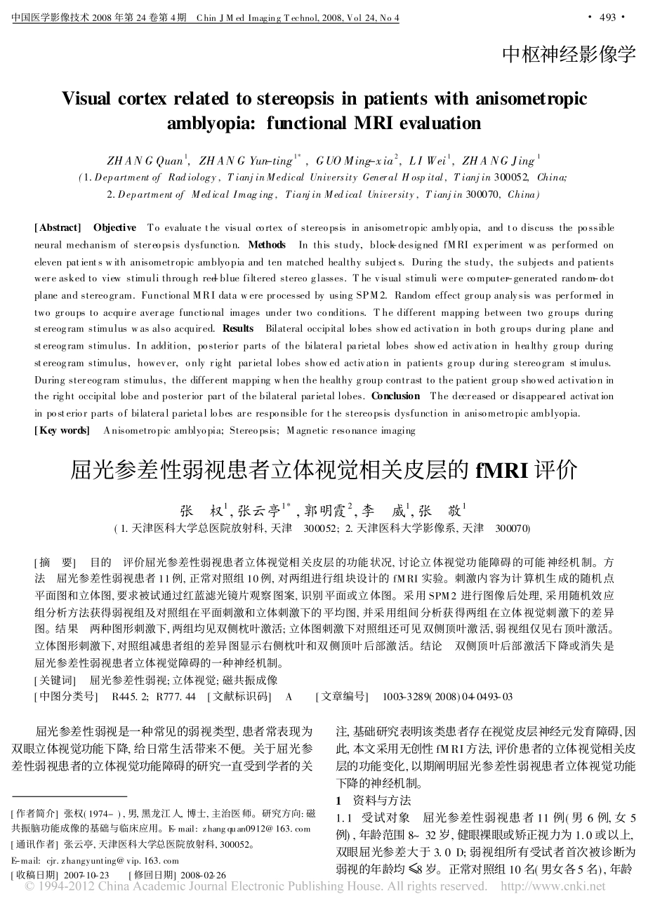
Duringthestudy,thesubjectsandpatientswereaskedtoviewstimulithroughred-bluefilteredstereoglasses.Thevisualstimuliwerecomputer-generatedrandom-dotplaneandstereogram.FunctionalMRIdatawereprocessedbyusingSPM2.Randomeffectgroupanalysiswasperformedintwogroupstoacquireaveragefunctionalimagesundertwoconditions.Thedifferentmappingbetweentwogroupsduringstereogramstimuluswasalsoacquired.ResultsBilateraloccipitallobesshowedactivationinbothgroupsduringplaneandstereogramstimulus.Inaddition,posteriorpartsofthebilateralparietallobesshowedactivationinhealthygroupduringstereogramstimulus,however,onlyrightparietallobesshowedactivationinpatientsgroupduringstereogramstimulus.Duringstereogramstimulus,thedifferentmappingwhenthehealthygroupcontrasttothepatientgroupshowedactivationintherightoccipitallobeandposteriorpartofthebilateralparietallobes.ConclusionThedecreasedordisappearedactivationinposteriorpartsofbilateralparietallobesareresponsibleforthestereopsisdysfunctioninanisometropicamblyopia.[Keywords]Anisometropicamblyopia;Stereopsis;Magneticresonanceimaging[作者简介]张权(1974-),男,黑龙江人,博士,主治医师。研究方向:磁共振脑功能成像的基础与临床应用。E-mail:zhangquan0912@163.com[通讯作者]张云亭,天津医科大学总医院放射科,300052。E-mail:cjr.zhangyunting@vip.163.com[收稿日期]2007-10-23[修回日期]2008-02-26屈光参差性弱视患者立体视觉相关皮层的fMRI评价张权1,张云亭1*,郭明霞2,李威1,张敬1(1.天津医科大学总医院放射科,天津300052;2.天津医科大学影像系,天津300070)[摘要]目的评价屈光参差性弱视患者立体视觉相关皮层的功能状况,讨论立体视觉功能障碍的可能神经机制。方法屈光参差性弱视患者11例,正常对照组10例,对两组进行组块设计的fMRI实验。刺激内容为计算机生成的随机点平面图和立体图,要求被试通过红蓝滤光镜片观察图案,识别平面或立体图。采用SPM2进行图像后处理,采用随机效应组分析方法获得弱视组及对照组在平面刺激和立体刺激下的平均图,并采用组间分析获得两组在立体视觉刺激下的差异图。结果两种图形刺激下,两组均见双侧枕叶激活;立体图刺激下对照组还可见双侧顶叶激活,弱视组仅见右顶叶激活。立体图形刺激下,对照组减患者组的差异图显示右侧枕叶和双侧顶叶后部激活。结论双侧顶叶后部激活下降或消失是屈光参差性弱视患者立体视觉障碍的一种神经机制。[关键词]屈光参差性弱视;立体视觉;磁共振成像[中图分类号]R445.2;R777.44[文献标识码]A[文章编号]1003-3289(2008)04-0493-03屈光参差性弱视是一种常见的弱视类型,患者常表现为双眼立体视觉功能下降,给日常生活带来不便。关于屈光参差性弱视患者的立体视觉功能障碍的研究一直受到学者的关注,基础研究表明该类患者存在视觉皮层神经元发育障碍,因此,本文采用无创性fMRI方法,评价患者的立体视觉相关皮层的功能变化,以期阐明屈光参差性弱视患者立体视觉功能下降的神经机制。1资料与方法1.1受试对象屈光参差性弱视患者11例(男6例,女5例),年龄范围8~32岁,健眼裸眼或矫正视力为1.0或以上,双眼屈光参差大于3.0D;弱视组所有受试者首次被诊断为弱视的年龄均[8岁。正常对照组10名(男女各5名),年龄#493#...